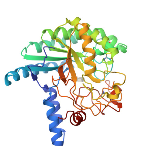
Activity and X-ray crystal structure of candidate base catalyst mutants of glycoside hydrolase family 6 cellobiohydrolase
Yamaguchi, S., Sunagawa, N., Tachioka, M., Igarashi, K.To be published.
Experimental Data Snapshot
Starting Model: experimental
View more details
Entity ID: 1 | |||||
|---|---|---|---|---|---|
| Molecule | Chains | Sequence Length | Organism | Details | Image |
| Glucanase | 358 | Phanerodontia chrysosporium | Mutation(s): 2 Gene Names: cel6A EC: 3.2.1 | 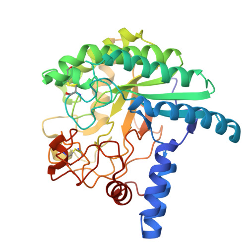 | |
UniProt | |||||
Find proteins for Q02321 (Phanerodontia chrysosporium) Explore Q02321 Go to UniProtKB: Q02321 | |||||
Entity Groups | |||||
| Sequence Clusters | 30% Identity50% Identity70% Identity90% Identity95% Identity100% Identity | ||||
| UniProt Group | Q02321 | ||||
Sequence AnnotationsExpand | |||||
| |||||
| Ligands 1 Unique | |||||
|---|---|---|---|---|---|
| ID | Chains | Name / Formula / InChI Key | 2D Diagram | 3D Interactions | |
| IDC (Subject of Investigation/LOI) Query on IDC | B [auth A] | (5R,6R,7R,8S)-7,8-dihydroxy-5-(hydroxymethyl)-5,6,7,8-tetrahydroimidazo[1,2-a]pyridin-6-yl beta-D-glucopyranoside C14 H22 N2 O9 CSXOUJBOYXGFCL-OFKZETBZSA-N |  | ||
| Length ( Å ) | Angle ( ˚ ) |
|---|---|
| a = 54.825 | α = 90 |
| b = 67.239 | β = 90 |
| c = 88.36 | γ = 90 |
| Software Name | Purpose |
|---|---|
| PHENIX | refinement |
| XDS | data reduction |
| XDS | data scaling |
| PHASER | phasing |
| Funding Organization | Location | Grant Number |
|---|---|---|
| Japan Society for the Promotion of Science (JSPS) | Japan | 22J12651 |
RCSB PDB is hosted by

RCSB PDB is a member of the