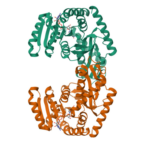
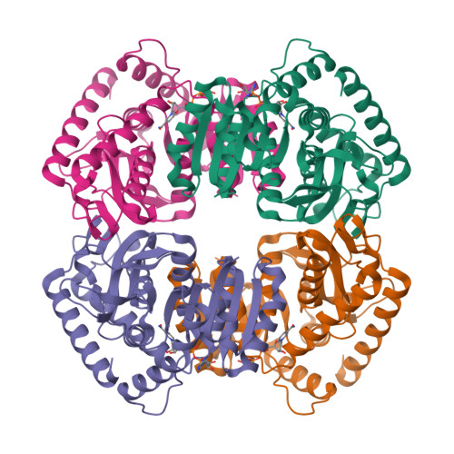
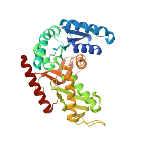
1LLD | pdb_00001lld

MOLECULAR BASIS OF ALLOSTERIC ACTIVATION OF BACTERIAL L-LACTATE DEHYDROGENASE


Experimental Data Snapshot

  • Method: X-RAY DIFFRACTION
  • Resolution: 2.00 Å
  • R-Value Work: 
    0.170 (DCC) 
  • R-Value Observed: 
    0.179 (Depositor) 

wwPDB Validation   3D Report Full Report


Ligand Structure Quality Assessment 

Created with Raphaël 2.3.0Worse 01 BetterLigand structure goodness of fit to experimental dataBest fitted NADClick on this verticalbar to view details

This is version 1.4 of the entry. See complete history


Literature

Molecular basis of allosteric activation of bacterial L-lactate dehydrogenase.

Iwata, S.Ohta, T.

(1993) J Mol Biology 230: 21-27

  • DOI: https://doi.org/10.1006/jmbi.1993.1122
  • Primary Citation of Related Structures:  
    1LLD

  • PubMed Abstract: 

    The three-dimensional structure of allosteric L-lactate dehydrogenase from Bifidobacterium longum, the first example of a T-state structure of L-lactate dehydrogenase, has been determined to 2.0 A. A comparative study of this structure with the previously reported R-state structure from Bacillus stearothermophilus has revealed the allosteric activation mechanism of the bacterial L-lactate dehydrogenase. The fructose 1,6-bisphosphate-induced conformational change at the effector site and the substrate affinity change at the activity site are clearly shown at a molecular level. Coupling of these changes can be simply explained by a set of concerted rotations between subunits in the tetramer of the enzyme. This T to R transition is the first example for a tetrameric allosteric protein where the rotations occur around each of three axes of symmetry.


  • Organizational Affiliation

    Photon Factory, National Laboratory for High Energy Physics, Ibaraki-ken, Japan.


Macromolecules
Find similar proteins by:  (by identity cutoff)  |  3D Structure
Entity ID: 1
MoleculeChains Sequence LengthOrganismDetailsImage
L-LACTATE DEHYDROGENASE
A, B
319Bifidobacterium longum subsp. longumMutation(s): 0 
EC: 1.1.1.27
UniProt
Find proteins for E8ME30 (Bifidobacterium longum subsp. longum (strain ATCC 15707 / DSM 20219 / JCM 1217 / NCTC 11818 / E194b))
Explore E8ME30 
Go to UniProtKB:  E8ME30
Entity Groups  
Sequence Clusters30% Identity50% Identity70% Identity90% Identity95% Identity100% Identity
UniProt GroupE8ME30
Sequence Annotations
Expand
  • Reference Sequence
Experimental Data & Validation

Experimental Data

  • Method: X-RAY DIFFRACTION
  • Resolution: 2.00 Å
  • R-Value Work:  0.170 (DCC) 
  • R-Value Observed: 0.179 (Depositor) 
Space Group: P 21 21 2
Unit Cell:
Length ( Å )Angle ( ˚ )
a = 105.8α = 90
b = 131.4β = 90
c = 63.8γ = 90
Software Package:
Software NamePurpose
PROLSQrefinement

Structure Validation

View Full Validation Report


Ligand Structure Quality Assessment 

Created with Raphaël 2.3.0Worse 01 BetterLigand structure goodness of fit to experimental dataBest fitted NADClick on this verticalbar to view details

Entry History 

Deposition Data

Revision History  (Full details and data files)

  • Version 1.0: 1994-01-31
    Type: Initial release
  • Version 1.1: 2008-03-03
    Changes: Version format compliance
  • Version 1.2: 2011-07-13
    Changes: Derived calculations, Version format compliance
  • Version 1.3: 2017-11-29
    Changes: Derived calculations, Other
  • Version 1.4: 2024-02-14
    Changes: Data collection, Database references, Derived calculations