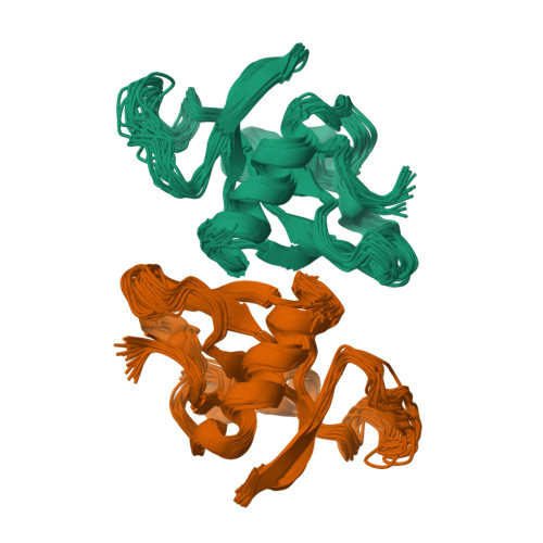
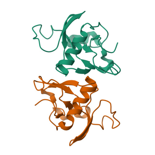
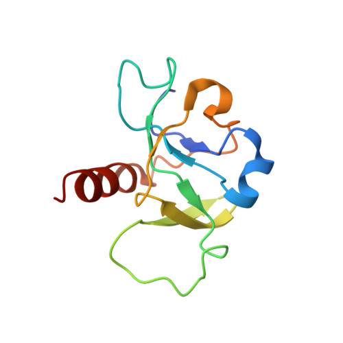
Structural and Thermodynamic Characterization of Vibrio fischeri CcdB.
De Jonge, N., Hohlweg, W., Garcia-Pino, A., Respondek, M., Buts, L., Haesaerts, S., Lah, J., Zangger, K., Loris, R.(2010) J Biological Chem 285: 5606-5613
- PubMed: 19959472
- DOI: https://doi.org/10.1074/jbc.M109.068429
- Primary Citation of Related Structures:
2KMT, 3JRZ, 3JSC - PubMed Abstract:
CcdB(Vfi) from Vibrio fischeri is a member of the CcdB family of toxins that poison covalent gyrase-DNA complexes. In solution CcdB(Vfi) is a dimer that unfolds to the corresponding monomeric components in a two-state fashion. In the unfolded state, the monomer retains a partial secondary structure. This observation correlates well with the crystal and NMR structures of the protein, which show a dimer with a hydrophobic core crossing the dimer interface. In contrast to its F plasmid homologue, CcdB(Vfi) possesses a rigid dimer interface, and the apparent relative rotations of the two subunits are due to structural plasticity of the monomer. CcdB(Vfi) shows a number of non-conservative substitutions compared with the F plasmid protein in both the CcdA and the gyrase binding sites. Although variation in the CcdA interaction site likely determines toxin-antitoxin specificity, substitutions in the gyrase-interacting region may have more profound functional implications.
Organizational Affiliation:
Structural Biology Brussels, and Department of Molecular and Cellular Interactions, Vrije Universiteit Brussel, Pleinlaan 2, B-1050 Brussels, Belgium.