4D59 | pdb_00004d59

Clostridial Cysteine protease Cwp84 C116A after propeptide cleavage


Experimental Data Snapshot

  • Method: X-RAY DIFFRACTION
  • Resolution: 1.84 Å
  • R-Value Free: 
    0.291 (Depositor), 0.300 (DCC) 
  • R-Value Work: 
    0.227 (Depositor), 0.230 (DCC) 
  • R-Value Observed: 
    0.230 (Depositor) 

Starting Model: experimental
View more details

wwPDB Validation   3D Report Full Report


Ligand Structure Quality Assessment 

Created with Raphaël 2.3.0Worse 01 BetterLigand structure goodness of fit to experimental dataBest fitted JEFClick on this verticalbar to view details

This is version 1.3 of the entry. See complete history


Literature

Cwp84, a Clostridium Difficile Cysteine Protease, Exhibits Conformational Flexibility in the Absence of its Propeptide

Bradshaw, W.J.Roberts, A.K.Shone, C.C.Acharya, K.R.

(2015) Acta Crystallogr Sect F Struct Biol Cryst Commun 71: 295

  • DOI: https://doi.org/10.1107/S2053230X15001065
  • Primary Citation of Related Structures:  
    4D59, 4D5A

  • PubMed Abstract: 

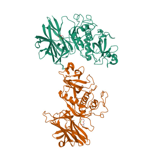
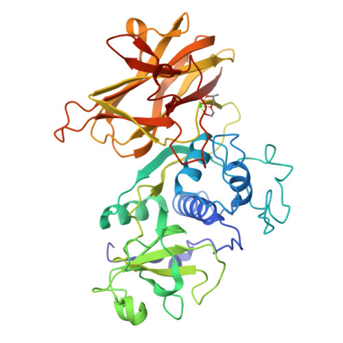
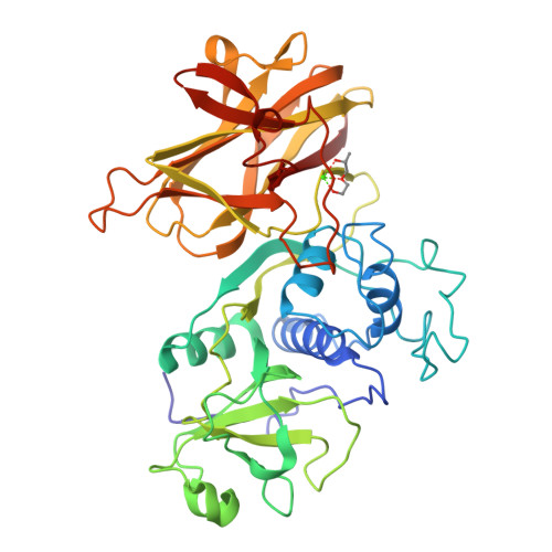
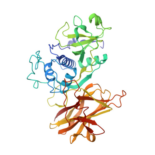
    In recent decades, the global healthcare problems caused by Clostridium difficile have increased at an alarming rate. A greater understanding of this antibiotic-resistant bacterium, particularly with respect to how it interacts with the host, is required for the development of novel strategies for fighting C. difficile infections. The surface layer (S-layer) of C. difficile is likely to be of significant importance to host-pathogen interactions. The mature S-layer is formed by a proteinaceous array consisting of multiple copies of a high-molecular-weight and a low-molecular-weight S-layer protein. These components result from the cleavage of SlpA by Cwp84, a cysteine protease. The structure of a truncated Cwp84 active-site mutant has recently been reported and the key features have been identified, providing the first structural insights into the role of Cwp84 in the formation of the S-layer. Here, two structures of Cwp84 after propeptide cleavage are presented and the three conformational changes that are observed are discussed. These changes result in a reconfiguration of the active site and exposure of the hydrophobic pocket.


  • Organizational Affiliation

    Department of Biology and Biochemistry, University of Bath, Claverton Down, Bath BA2 7AY, England.


Macromolecules
Find similar proteins by:  (by identity cutoff)  |  3D Structure
Entity ID: 1
MoleculeChains Sequence LengthOrganismDetailsImage
CELL SURFACE PROTEIN (PUTATIVE CELL SURFACE-ASSOCIATED CYSTEINE PROTEASE)
A, B
406Clostridioides difficile QCD-32g58Mutation(s): 1 
EC: 3.4.22.15
Entity Groups  
Sequence Clusters30% Identity50% Identity70% Identity90% Identity95% Identity100% Identity
Sequence Annotations
Expand
  • Reference Sequence
Experimental Data & Validation

Experimental Data

  • Method: X-RAY DIFFRACTION
  • Resolution: 1.84 Å
  • R-Value Free:  0.291 (Depositor), 0.300 (DCC) 
  • R-Value Work:  0.227 (Depositor), 0.230 (DCC) 
  • R-Value Observed: 0.230 (Depositor) 
Space Group: P 1
Unit Cell:
Length ( Å )Angle ( ˚ )
a = 42.22α = 89.28
b = 58.421β = 77.96
c = 93.1γ = 71.62
Software Package:
Software NamePurpose
REFMACrefinement
XDSdata reduction
xia2data scaling
PHASERphasing

Structure Validation

View Full Validation Report


Ligand Structure Quality Assessment 

Created with Raphaël 2.3.0Worse 01 BetterLigand structure goodness of fit to experimental dataBest fitted JEFClick on this verticalbar to view details

Entry History 

Deposition Data

Revision History  (Full details and data files)

  • Version 1.0: 2015-03-04
    Type: Initial release
  • Version 1.1: 2015-03-11
    Changes: Database references
  • Version 1.2: 2015-03-25
    Changes: Database references
  • Version 1.3: 2023-12-20
    Changes: Advisory, Data collection, Database references, Derived calculations, Other, Refinement description