4PNJ | pdb_00004pnj

Recombinant Sperm Whale P6 Myoglobin Solved with Single Pulse Free Electron Laser Data


Experimental Data Snapshot

  • Method: X-RAY DIFFRACTION
  • Resolution: 1.36 Å
  • R-Value Free: 
    0.188 (Depositor), 0.190 (DCC) 
  • R-Value Work: 
    0.159 (Depositor), 0.160 (DCC) 
  • R-Value Observed: 
    0.160 (Depositor) 

Starting Model: experimental
View more details

wwPDB Validation   3D Report Full Report


Ligand Structure Quality Assessment 

Created with Raphaël 2.3.0Worse 01 BetterLigand structure goodness of fit to experimental dataBest fitted HEMClick on this verticalbar to view details

This is version 1.7 of the entry. See complete history


Literature

Goniometer-based femtosecond crystallography with X-ray free electron lasers.

Cohen, A.E.Soltis, S.M.Gonzalez, A.Aguila, L.Alonso-Mori, R.Barnes, C.O.Baxter, E.L.Brehmer, W.Brewster, A.S.Brunger, A.T.Calero, G.Chang, J.F.Chollet, M.Ehrensberger, P.Eriksson, T.L.Feng, Y.Hattne, J.Hedman, B.Hollenbeck, M.Holton, J.M.Keable, S.Kobilka, B.K.Kovaleva, E.G.Kruse, A.C.Lemke, H.T.Lin, G.Lyubimov, A.Y.Manglik, A.Mathews, I.I.McPhillips, S.E.Nelson, S.Peters, J.W.Sauter, N.K.Smith, C.A.Song, J.Stevenson, H.P.Tsai, Y.Uervirojnangkoorn, M.Vinetsky, V.Wakatsuki, S.Weis, W.I.Zadvornyy, O.A.Zeldin, O.B.Zhu, D.Hodgson, K.O.

(2014) Proc Natl Acad Sci U S A 111: 17122-17127

  • DOI: https://doi.org/10.1073/pnas.1418733111
  • Primary Citation of Related Structures:  
    4PNJ

  • PubMed Abstract: 

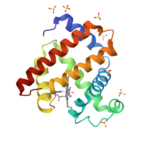
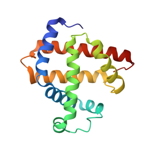
    The emerging method of femtosecond crystallography (FX) may extend the diffraction resolution accessible from small radiation-sensitive crystals and provides a means to determine catalytically accurate structures of acutely radiation-sensitive metalloenzymes. Automated goniometer-based instrumentation developed for use at the Linac Coherent Light Source enabled efficient and flexible FX experiments to be performed on a variety of sample types. In the case of rod-shaped Cpl hydrogenase crystals, only five crystals and about 30 min of beam time were used to obtain the 125 still diffraction patterns used to produce a 1.6-Å resolution electron density map. For smaller crystals, high-density grids were used to increase sample throughput; 930 myoglobin crystals mounted at random orientation inside 32 grids were exposed, demonstrating the utility of this approach. Screening results from cryocooled crystals of β2-adrenoreceptor and an RNA polymerase II complex indicate the potential to extend the diffraction resolution obtainable from very radiation-sensitive samples beyond that possible with undulator-based synchrotron sources.


  • Organizational Affiliation

    Stanford Synchrotron Radiation Lightsource, HodgsonK@stanford.edu acohen@slac.stanford.edu.


Macromolecules
Find similar proteins by:  (by identity cutoff)  |  3D Structure
Entity ID: 1
MoleculeChains Sequence LengthOrganismDetailsImage
Myoglobin154Physeter macrocephalusMutation(s): 0 
Gene Names: MB
EC: 1.11.1 (UniProt), 1.7 (UniProt)
UniProt
Find proteins for P02185 (Physeter macrocephalus)
Explore P02185 
Go to UniProtKB:  P02185
Entity Groups  
Sequence Clusters30% Identity50% Identity70% Identity90% Identity95% Identity100% Identity
UniProt GroupP02185
Sequence Annotations
Expand
  • Reference Sequence
Small Molecules
Ligands 2 Unique
IDChains Name / Formula / InChI Key2D Diagram3D Interactions
HEM
Query on HEM

Download Ideal Coordinates CCD File 
B [auth A]PROTOPORPHYRIN IX CONTAINING FE
C34 H32 Fe N4 O4
KABFMIBPWCXCRK-RGGAHWMASA-L
SO4
Query on SO4

Download Ideal Coordinates CCD File 
C [auth A]
D [auth A]
E [auth A]
F [auth A]
G [auth A]
SULFATE ION
O4 S
QAOWNCQODCNURD-UHFFFAOYSA-L
Experimental Data & Validation

Experimental Data

  • Method: X-RAY DIFFRACTION
  • Resolution: 1.36 Å
  • R-Value Free:  0.188 (Depositor), 0.190 (DCC) 
  • R-Value Work:  0.159 (Depositor), 0.160 (DCC) 
  • R-Value Observed: 0.160 (Depositor) 
Space Group: P 6
Unit Cell:
Length ( Å )Angle ( ˚ )
a = 90.309α = 90
b = 90.309β = 90
c = 45.204γ = 120
Software Package:
Software NamePurpose
cctbx.xfeldata reduction
MOLREPphasing
PDB_EXTRACTdata extraction
PHENIXrefinement

Structure Validation

View Full Validation Report


Ligand Structure Quality Assessment 

Created with Raphaël 2.3.0Worse 01 BetterLigand structure goodness of fit to experimental dataBest fitted HEMClick on this verticalbar to view details

Entry History 

Deposition Data

Revision History  (Full details and data files)

  • Version 1.0: 2014-11-05
    Type: Initial release
  • Version 1.1: 2014-12-03
    Changes: Database references
  • Version 1.2: 2014-12-17
    Changes: Database references
  • Version 1.3: 2015-04-01
    Changes: Other
  • Version 1.4: 2015-11-18
    Changes: Data collection
  • Version 1.5: 2017-11-15
    Changes: Data collection, Derived calculations, Refinement description
  • Version 1.6: 2018-11-28
    Changes: Data collection
  • Version 1.7: 2023-09-27
    Changes: Data collection, Database references, Refinement description