7JRH

X-ray crystal structure of a cyclic peptide containing medin(19-25) and medin(31-37)

  • Classification: DE NOVO PROTEIN
  • Organism(s): Homo sapiens
  • Mutation(s): No 

  • Deposited: 2020-08-12 Released: 2020-09-09 
  • Deposition Author(s): Wierzbicki, M., Howitz, W.J., Nowick, J.S.
  • Funding Organization(s): National Institutes of Health/National Institute of General Medical Sciences (NIH/NIGMS), National Science Foundation (NSF, United States)

Experimental Data Snapshot

  • Method: X-RAY DIFFRACTION
  • Resolution: 1.32 Å
  • R-Value Free: 0.210 
  • R-Value Work: 0.177 
  • R-Value Observed: 0.181 

wwPDB Validation   3D Report Full Report


This is version 1.1 of the entry. See complete history


Literature

Interpenetrating Cubes in the X-ray Crystallographic Structure of a Peptide Derived from Medin 19-36 .

Howitz, W.J.Wierzbicki, M.Cabanela, R.W.Saliba, C.Motavalli, A.Tran, N.Nowick, J.S.

(2020) J Am Chem Soc 142: 15870-15875

  • DOI: https://doi.org/10.1021/jacs.0c06143
  • Primary Citation of Related Structures:  
    7JRH

  • PubMed Abstract: 

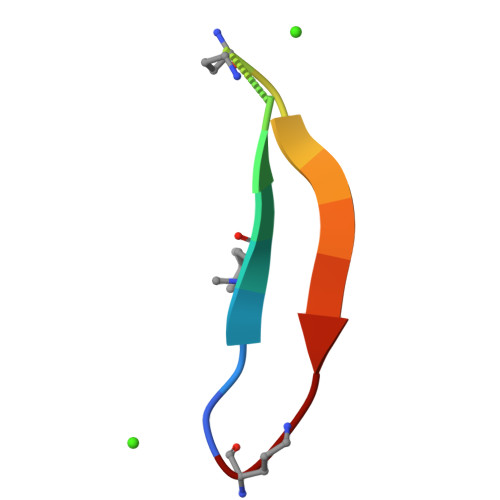
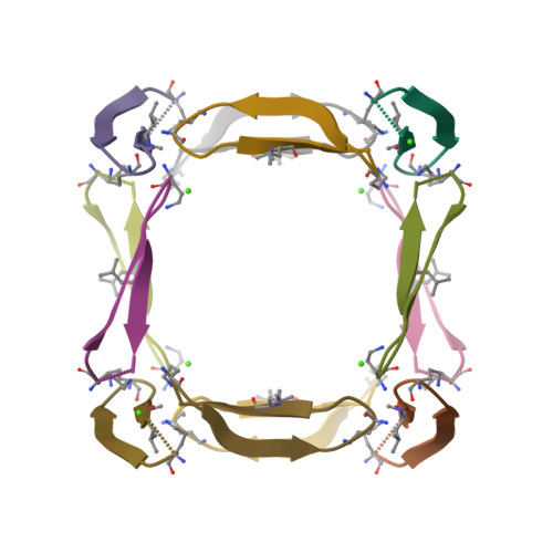
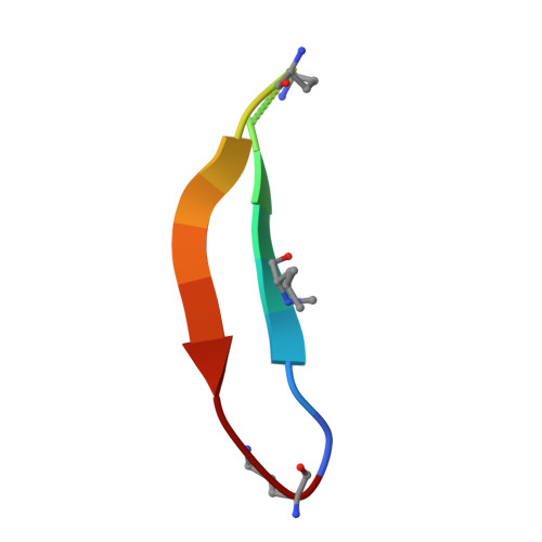
    Amyloidogenic peptides and proteins are rich sources of supramolecular assemblies. Sequences derived from well-known amyloids, including Aβ, human islet amyloid polypeptide, and tau have been found to assemble as fibrils, nanosheets, ribbons, and nanotubes. The supramolecular assembly of medin, a 50-amino acid peptide that forms fibrillary deposits in aging human vasculature, has not been heavily investigated. In this work, we present an X-ray crystallographic structure of a cyclic β-sheet peptide derived from the 19-36 region of medin that assembles to form interpenetrating cubes. The edge of each cube is composed of a single peptide, and each vertex is occupied by a divalent metal ion. This structure may be considered a metal-organic framework (MOF) containing a large peptide ligand. This work demonstrates that peptides containing Glu or Asp that are preorganized to adopt β-hairpin structures can serve as ligands and assemble with metal ions to form MOFs.


  • Organizational Affiliation

    Department of Chemistry and Department of Pharmaceutical Sciences, University of California, Irvine, Irvine, California 92697-2025, United States.


Macromolecules

Find similar proteins by:  Sequence   |   3D Structure  

Entity ID: 1
MoleculeChains Sequence LengthOrganismDetailsImage
Cyclic peptide ASP-GLN-TRP-MLE-GLN-VAL-ASP-ORD-GLU-VAL-THR-GLY-ILE-ILE-THR-ORD16Homo sapiensMutation(s): 0 
Entity Groups  
Sequence Clusters30% Identity50% Identity70% Identity90% Identity95% Identity100% Identity
Sequence Annotations
Expand
  • Reference Sequence
Small Molecules
Modified Residues  2 Unique
IDChains TypeFormula2D DiagramParent
MLE
Query on MLE
A
L-PEPTIDE LINKINGC7 H15 N O2LEU
ORN
Query on ORN
A
L-PEPTIDE LINKINGC5 H12 N2 O2ALA
Experimental Data & Validation

Experimental Data

  • Method: X-RAY DIFFRACTION
  • Resolution: 1.32 Å
  • R-Value Free: 0.210 
  • R-Value Work: 0.177 
  • R-Value Observed: 0.181 
  • Space Group: I 2 3
Unit Cell:
Length ( Å )Angle ( ˚ )
a = 42.486α = 90
b = 42.486β = 90
c = 42.486γ = 90
Software Package:
Software NamePurpose
PHENIXrefinement
SAINTdata reduction
SADABSdata scaling
AutoSolphasing

Structure Validation

View Full Validation Report


Entry History & Funding Information

Deposition Data


Funding OrganizationLocationGrant Number
National Institutes of Health/National Institute of General Medical Sciences (NIH/NIGMS)United StatesGM097562
National Science Foundation (NSF, United States)United StatesCHE1808096

Revision History  (Full details and data files)

  • Version 1.0: 2020-09-09
    Type: Initial release
  • Version 1.1: 2020-09-30
    Changes: Database references