8TB2

Structure of SasG (type II) (residues 165-421) from Staphylococcus aureus MW2


Experimental Data Snapshot

  • Method: X-RAY DIFFRACTION
  • Resolution: 1.88 Å
  • R-Value Free: 0.253 
  • R-Value Work: 0.184 

wwPDB Validation   3D Report Full Report


This is version 1.0 of the entry. See complete history


Literature

Staphylococcus aureus skin colonization is mediated by SasG lectin variation.

Mills, K.B.Maciag, J.J.Wang, C.Crawford, J.A.Enroth, T.J.Keim, K.C.Dufrene, Y.F.Robinson, D.A.Fey, P.D.Herr, A.B.Horswill, A.R.

(2024) Cell Rep 43: 114022-114022

  • DOI: https://doi.org/10.1016/j.celrep.2024.114022
  • Primary Citation of Related Structures:  
    8TB2

  • PubMed Abstract: 

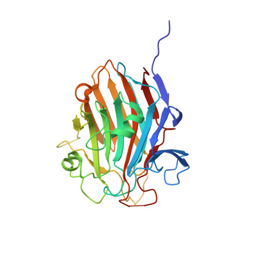
    Staphylococcus aureus causes the majority of skin and soft tissue infections, but this pathogen only transiently colonizes healthy skin. However, this transient skin exposure enables S. aureus to transition to infection. The initial adhesion of S. aureus to skin corneocytes is mediated by surface protein G (SasG). Here, phylogenetic analyses reveal the presence of two major divergent SasG alleles in S. aureus: SasG-I and SasG-II. Structural analyses of SasG-II identify a nonaromatic arginine in the binding pocket of the lectin subdomain that mediates adhesion to corneocytes. Atomic force microscopy and corneocyte adhesion assays indicate that SasG-II can bind to a broader variety of ligands than SasG-I. Glycosidase treatment results in different binding profiles between SasG-I and SasG-II on skin cells. In addition, SasG-mediated adhesion is recapitulated using differentiated N/TERT keratinocytes. Our findings indicate that SasG-II has evolved to adhere to multiple ligands, conferring a distinct advantage to S. aureus during skin colonization.


  • Organizational Affiliation

    Department of Immunology and Microbiology, University of Colorado Anschutz Medical Campus, Aurora, CO, USA.


Macromolecules
Find similar proteins by:  (by identity cutoff)  |  3D Structure
Entity ID: 1
MoleculeChains Sequence LengthOrganismDetailsImage
Putative surface protein MW2416262Staphylococcus aureus subsp. aureus MW2Mutation(s): 0 
Gene Names: MW2416
UniProt
Find proteins for Q8NUV0 (Staphylococcus aureus (strain MW2))
Explore Q8NUV0 
Go to UniProtKB:  Q8NUV0
Entity Groups  
Sequence Clusters30% Identity50% Identity70% Identity90% Identity95% Identity100% Identity
UniProt GroupQ8NUV0
Sequence Annotations
Expand
  • Reference Sequence
Small Molecules
Ligands 4 Unique
IDChains Name / Formula / InChI Key2D Diagram3D Interactions
SO4
Query on SO4

Download Ideal Coordinates CCD File 
B [auth A],
C [auth A],
D [auth A],
E [auth A]
SULFATE ION
O4 S
QAOWNCQODCNURD-UHFFFAOYSA-L
ZN
Query on ZN

Download Ideal Coordinates CCD File 
I [auth A],
J [auth A],
K [auth A]
ZINC ION
Zn
PTFCDOFLOPIGGS-UHFFFAOYSA-N
CA (Subject of Investigation/LOI)
Query on CA

Download Ideal Coordinates CCD File 
F [auth A],
G [auth A]
CALCIUM ION
Ca
BHPQYMZQTOCNFJ-UHFFFAOYSA-N
NA
Query on NA

Download Ideal Coordinates CCD File 
H [auth A]SODIUM ION
Na
FKNQFGJONOIPTF-UHFFFAOYSA-N
Experimental Data & Validation

Experimental Data

  • Method: X-RAY DIFFRACTION
  • Resolution: 1.88 Å
  • R-Value Free: 0.253 
  • R-Value Work: 0.184 
  • Space Group: P 21 21 2
Unit Cell:
Length ( Å )Angle ( ˚ )
a = 78.6α = 90
b = 70.21β = 90
c = 43.54γ = 90
Software Package:
Software NamePurpose
PHENIXrefinement
MOSFLMdata reduction
Aimlessdata scaling
PHASERphasing

Structure Validation

View Full Validation Report


Entry History & Funding Information

Deposition Data


Funding OrganizationLocationGrant Number
National Institutes of Health/National Institute of General Medical Sciences (NIH/NIGMS)United StatesR01 GM094363
National Institutes of Health/National Institute Of Allergy and Infectious Diseases (NIH/NIAID)United StatesR01 AI162964

Revision History  (Full details and data files)

  • Version 1.0: 2024-05-08
    Type: Initial release