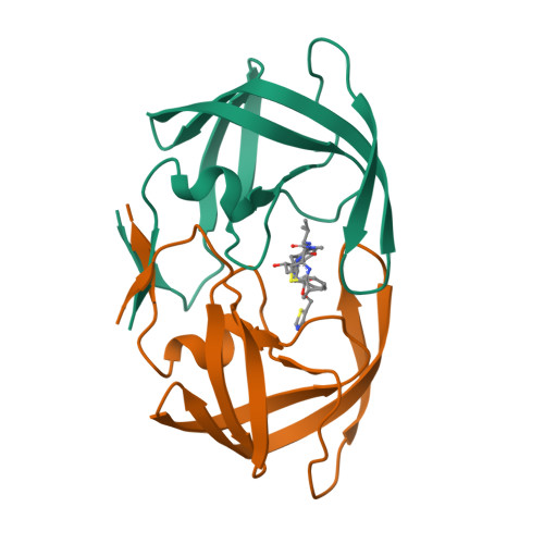
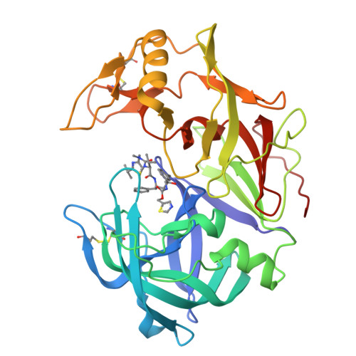
Structure Determination MethodologyScientific Name of Source OrganismRefinement Resolution (Å)Enzyme Classification NameMembrane Protein Annotation | Rezacova, P., Brynda, J., Sedlacek, J., Konvalinka, J., Fabry, M., Horejsi, M. (2005) To be published | Released | 2005-04-19 | | Method | X-RAY DIFFRACTION 2 Å | | Organisms | | | Macromolecule | | | Unique Ligands | RIT |
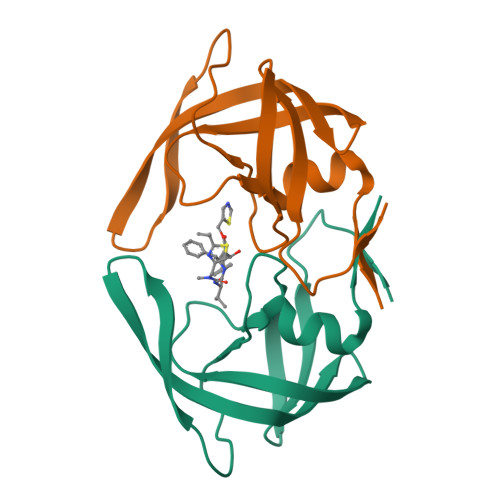
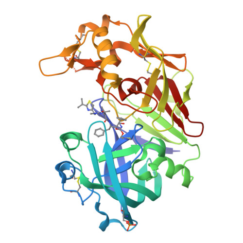
Clemente, J.C., Moose, R.E., Hemrajani, R., Govindasamy, L., Reutzel, R., McKenna, R., Abanje-McKenna, M., Goodenow, M.M., Dunn, B.M. (2004) Biochemistry 43: 12141-12151 | Released | 2004-10-05 | | Method | X-RAY DIFFRACTION 2.5 Å | | Organisms | | | Macromolecule | | | Unique Ligands | RIT |
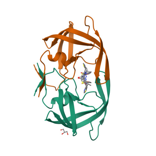
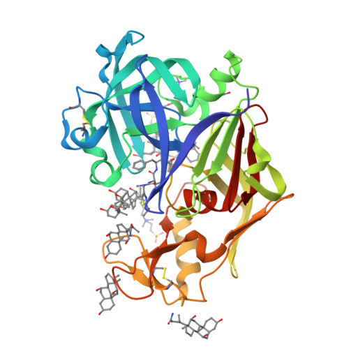
Clemente, J.C., Stow, L.R., Janka, L.K., Jeung, J.A., Desai, K.A., Govindasamy, L., Agbandje-McKenna, M., McKenna, R., Goodenow, M.M., Dunn, B.M. To be published | Released | 2006-11-14 | | Method | X-RAY DIFFRACTION 2.2 Å | | Organisms | | | Macromolecule | | | Unique Ligands | GOL, RIT |
Dostal, J., Brynda, J., Hruskova-Heidingsfeldova, O., Pachl, P., Pichova, I., Rezacova, P. (2012) J Enzyme Inhib Med Chem 27: 160-165 | Released | 2012-03-07 | | Method | X-RAY DIFFRACTION 2.4 Å | | Organisms | | | Macromolecule | | | Unique Ligands | RIT |
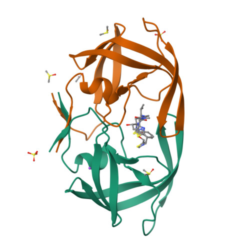
Liu, Z., Yedidi, R.S., Wang, Y., Brunzelle, J.S., Kovari, I.A., Kovari, L.C. (2013) Biochem Biophys Res Commun 431: 232-238 | Released | 2013-01-30 | | Method | X-RAY DIFFRACTION 1.8 Å | | Organisms | | | Macromolecule | | | Unique Ligands | RIT |
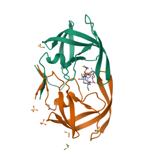
Yedidi, R.S., Garimella, H., Chang, S.B., Kaufman, J.D., Das, D., Wingfield, P.T., Mitsuya, H. (2014) Antimicrob Agents Chemother 58: 3679-3688 | Released | 2014-04-02 | | Method | X-RAY DIFFRACTION 1.8 Å | | Organisms | | | Macromolecule | | | Unique Ligands | RIT |
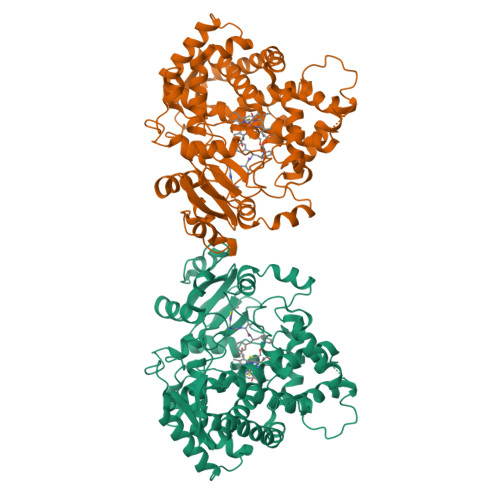
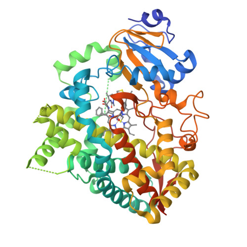
Sevrioukova, I. (2017) Biochemistry 56: 3058-3067 | Released | 2017-05-31 | | Method | X-RAY DIFFRACTION 2.7 Å | | Organisms | | | Macromolecule | | | Unique Ligands | HEM, RIT |
Sevrioukova, I. (2017) Biochemistry 56: 3058-3067 | Released | 2017-05-31 | | Method | X-RAY DIFFRACTION 2.2 Å | | Organisms | | | Macromolecule | | | Unique Ligands | HEM, RIT |
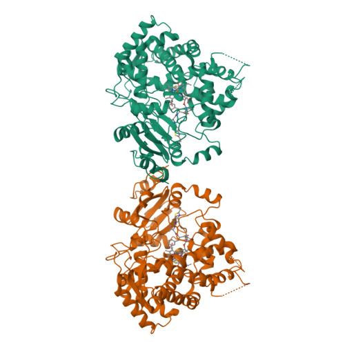
Hsu, M.-H., Johnson, E.F. (2018) Mol Pharmacol 93: 14-24 | Released | 2017-11-15 | | Method | X-RAY DIFFRACTION 2.91 Å | | Organisms | | | Macromolecule | | | Unique Ligands | HEM, RIT |
|