Warning You are using a web browser that we do not support. Our website will not work properly. Please update to a newer version or download a new web browser, such as Chrome or Firefox.
Help
Contact us
  • Sign in with Google
  • Sign in with Facebook
  • Sign in with ORCID

  • Help
RCSB PDB
  • Deposit 

      Prepare Data

    • PDBx/mmCIF file
    • pdb_extract
    • SF-Tool
    • Ligand Expo
    • MAXIT

      Validate Data

    • Validation Server
    • Validation API
    • Information for Journals
    • Validation Task Forces

      Deposit Data

    • wwPDB OneDep System
    • PDB-IHM

      Help and Resources

    • Deposit FAQ
    • Validation FAQ
    • Tutorials
    • Annotation Policies
    • Processing Procedures
    • PDBx/mmCIF Dictionary
    • PDBx/mmCIF User Guide
    • Chemical Component Dictionary
    • Biologically Interesting Molecule Reference Dictionary (BIRD)
    • BioSync/Beamlines/Facilities
    • Related Tools
  • Search 
    • Advanced Search
    • Sequence Similarity Search
    • Structure Similarity Search
    • Chemical Similarity Search
    • Chemical Sketch Tool
    • Browse by Annotations
    • New Entries
    • Unreleased Entries
    • PDB Statistics
  • Visualize 
    • Mol* (MolStar)
    • Sequence Annotations Viewer
    • Genome View
  • Analyze 
    • Pairwise Structure Alignment
    • Symmetry Resources in the PDB
    • Structure Quality
    • Grouping Structures
    • PDB Citation MeSH Network Explorer
    • PDB Statistics
    • EPPIC Biological Assemblies
    • External Data and Resources
    • Integrated Resources
    • Additional Resources
  • Download 
    • Coordinates and Experimental Data
    • Sequences
    • Ligands
    • File Download Services
    • Web APIs
  • Learn 

    • Training Courses
    • Guide to PDB Data
    • Molecule of the Month
    • Educational Resources
    • Curricula
    • Browse
    • News
    • SciArt Galleries
    • Irving Geis
    • David Goodsell
  • About 
    • Contact Us
    • About RCSB PDB
    • Vision and Mission
    • Citation, Usage, Privacy Policies, Logo
    • News
    • PDB History
    • PDB50
    • User Community
    • Publications
    • RCSB PDB Advisory Committee
    • Team Members
    • Service Status
  • Careers
  • COVID-19
RCSB PDB
244,730
Structures from the PDB archive
1,068,577
Computed Structure Models (CSM)
Include CSM
Advanced Search
|
Browse Annotations
Help
  • PDB-101
  • wwPDB
  • EMDataResource
  • NAKB: Nucleic Acid Knowledgebase
  • wwPDB Foundation
  • PDB-IHM Logo
  • Search
  • Query History
  • Browse Annotations
  • MyPDB
QUERY: BIRD Name HAS EXACT PHRASE "=beta-lactose"
MyPDB Login
 Search API
Advanced Search Query Builder
Help
Full Text
Structure Attributes
Chemical Attributes
Help
x
NOT
Count
Add Subquery
Sequence Similarity
Sequence Motif
Structure Similarity
Structure Motif
Chemical Similarity
Return
grouped by
Include Computed Structure Models (CSM)
Count
Clear
Search
Search Summary This query matches 146 Structures.
Refinements
Structure Determination Methodology
Scientific Name of Source Organism
More...
Taxonomy
Experimental Method
Polymer Entity Type
Refinement Resolution (Å)
Release Date
Enzyme Classification Name
Symmetry Type
SCOP Classification
1 to 25 of 146 Structures
Page 1 of 6
Sort by
Gallery item
 Explore in 3D

1AX1 | pdb_00001ax1

Download FileView File

ERYTHRINA CORALLODENDRON LECTIN IN COMPLEX WITH LACTOSE

Shaanan, B., Elgavish, S.

(1998) J Mol Biology 277: 917-932

Released1998-05-06
Method
X-RAY DIFFRACTION 1.95 Å
Organisms
Erythrina corallodendron
Macromolecule
LECTIN (protein)
Unique LigandsCA, MN
Unique branched monosaccharidesBGC, BMA, FUC, GAL, MAN, NAG, XYP
Gallery item
 Explore in 3D

1BZW | pdb_00001bzw

Download FileView File

PEANUT LECTIN COMPLEXED WITH C-LACTOSE

Ravishankar, R., Surolia, A., Vijayan, M., Lim, S., Kishi, Y.

(1998) J Am Chem Soc 120: 11297-11303
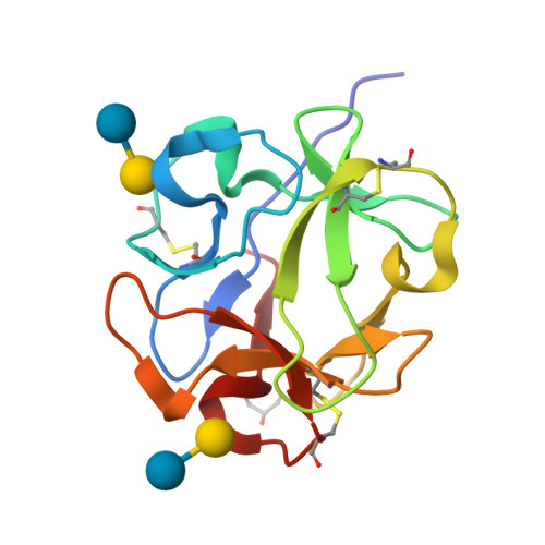
Released1998-11-11
Method
X-RAY DIFFRACTION 2.7 Å
Organisms
Arachis hypogaea
Macromolecule
PROTEIN (PEANUT LECTIN) (protein)
Unique LigandsCA, MN
Unique branched monosaccharidesBGC, GAL, GLC
Gallery item
 Explore in 3D

1C1L | pdb_00001c1l

Download FileView File

LACTOSE-LIGANDED CONGERIN I

Shirai, T., Mitsuyama, C., Niwa, Y., Matsui, Y., Hotta, H., Yamane, T., Kamiya, H., Ishii, C., Ogawa, T., Muramoto, K.

(1999) Structure 7: 1223-1233

Released1999-10-08
Method
X-RAY DIFFRACTION 1.5 Å
Organisms
Conger myriaster
Macromolecule
PROTEIN (CONGERIN I) (protein)
Unique branched monosaccharidesBGC, GAL
Gallery item
 Explore in 3D

1DLL | pdb_00001dll

Download FileView File

The HC fragement of tetanus toxin complexed with lactose

Emsley, P., Fotinou, C., Black, I., Fairweather, N.F., Charles, I.G., Watts, C., Hewitt, E., Isaacs, N.W.

(2000) J Biological Chem 275: 8889-8894

Released2000-03-24
Method
X-RAY DIFFRACTION 1.8 Å
Organisms
Clostridium tetani
Macromolecule
TETANUS TOXIN (protein)
Unique LigandsGOL
Unique branched monosaccharidesBGC, GAL
Gallery item
 Explore in 3D

1DVA | pdb_00001dva

Download FileView File

Crystal Structure of the Complex Between the Peptide Exosite Inhibitor E-76 and Coagulation Factor VIIA

Eigenbrot, C., Ultsch, M.H.

(2000) Nature 404: 465-470

Released2000-05-12
Method
X-RAY DIFFRACTION 3 Å
Organisms
Homo sapiens
Macromolecule
DES-GLA FACTOR VIIA (HEAVY CHAIN) (protein)
DES-GLA FACTOR VIIA (LIGHT CHAIN) (protein)
PEPTIDE E-76 (protein)
Unique Ligands0Z6, CA, CAC, FUC, FUL, GLC
Unique branched monosaccharidesBGC, GAL
Gallery item
 Explore in 3D

1GWV | pdb_00001gwv

Download FileView File

ALPHA-,1,3 GALACTOSYLTRANSFERASE - LACTOSE COMPLEX

Boix, E., Zhang, Y., Swaminathan, G.J., Brew, K., Acharya, K.R.

(2002) J Biological Chem 277: 28310

Released2003-03-20
Method
X-RAY DIFFRACTION 2.5 Å
Organisms
Bos taurus
Macromolecule
N-ACETYLLACTOSAMINIDE ALPHA-1,3-GALACTOSYLTRANSFERASE (protein)
Unique LigandsMN, UDP
Unique branched monosaccharidesBGC, GAL
Gallery item
 Explore in 3D

1GZC | pdb_00001gzc

Download FileView File

High-Resolution crystal structure of Erythrina cristagalli lectin in complex with lactose

Svensson, C., Krengel, U.

(2002) J Mol Biology 321: 69

Released2002-06-21
Method
X-RAY DIFFRACTION 1.58 Å
Organisms
Erythrina crista-galli
Macromolecule
ERYTHRINA CRISTA-GALLI LECTIN (protein)
Unique LigandsCA, MN
Unique branched monosaccharidesBGC, GAL
Gallery item
 Explore in 3D

1GZW | pdb_00001gzw

Download FileView File

X-RAY CRYSTAL STRUCTURE OF HUMAN GALECTIN-1

Lopez-Lucendo, M.I.F., Gabius, H.J., Romero, A.

(2004) J Mol Biology 343: 957

Released2004-01-08
Method
X-RAY DIFFRACTION 1.7 Å
Organisms
Homo sapiens
Macromolecule
GALECTIN-1 (protein)
Unique LigandsBME, SO4
Unique branched monosaccharidesBGC, GAL
Gallery item
 Explore in 3D

1HLC | pdb_00001hlc

Download FileView File

X-RAY CRYSTAL STRUCTURE OF THE HUMAN DIMERIC S-LAC LECTIN, L-14-II, IN COMPLEX WITH LACTOSE AT 2.9 ANGSTROMS RESOLUTION

Lobsanov, Y.D., Gitt, M.A., Leffler, H., Barondes, S., Rini, J.M.

(1993) J Biological Chem 268: 27034-27038

Released1994-04-30
Method
X-RAY DIFFRACTION 2.9 Å
Organisms
Homo sapiens
Macromolecule
HUMAN LECTIN (protein)
Unique branched monosaccharidesBGC, GAL
Gallery item
 Explore in 3D

1HWP | pdb_00001hwp

Download FileView File

EBULIN COMPLEXED WITH PTEROIC ACID, TRIGONAL CRYSTAL FORM

Pascal, J.M., Day, P.J., Monzingo, A.F., Ernst, S.R., Robertus, J.D.

(2001) Proteins 43: 319-326

Released2001-01-24
Method
X-RAY DIFFRACTION 3.1 Å
Organisms
Sambucus ebulus
Macromolecule
EBULIN (protein)
Unique LigandsPT1
Unique branched monosaccharidesBGC, BMA, GAL, NAG
Gallery item
 Explore in 3D

1IS3 | pdb_00001is3

Download FileView File

LACTOSE AND MES-LIGANDED CONGERIN II

Shirai, T., Matsui, Y., Shionyu-Mitsuyama, C., Yamane, T., Kamiya, H., Ishii, C., Ogawa, T., Muramoto, K.

(2002) J Mol Biology 321: 879-889

Released2002-09-18
Method
X-RAY DIFFRACTION 1.45 Å
Organisms
Conger myriaster
Macromolecule
CONGERIN II (protein)
Unique LigandsMES
Unique branched monosaccharidesBGC, GAL
Gallery item
 Explore in 3D

1IS4 | pdb_00001is4

Download FileView File

LACTOSE-LIGANDED CONGERIN II

Shirai, T., Matsui, Y., Shionyu-Mitsuyama, C., Yamane, T., Kamiya, H., Ishii, C., Ogawa, T., Muramoto, K.

(2002) J Mol Biology 321: 879-889

Released2002-09-18
Method
X-RAY DIFFRACTION 1.9 Å
Organisms
Conger myriaster
Macromolecule
CONGERIN II (protein)
Unique branched monosaccharidesBGC, GAL
Gallery item
 Explore in 3D

1IT0 | pdb_00001it0

Download FileView File

Crystal structure of xylanase from Streptomyces olivaceoviridis E-86 complexed with lactose

Fujimoto, Z., Kuno, A., Kaneko, S., Kobayashi, H., Kusakabe, I., Mizuno, H.

(2002) J Mol Biology 316: 65-78

Released2002-02-20
Method
X-RAY DIFFRACTION 2 Å
Organisms
Streptomyces olivaceoviridis
Macromolecule
endo-1,4-beta-D-xylanase (protein)
Unique branched monosaccharidesBGC, GAL
Gallery item
 Explore in 3D

1JYN | pdb_00001jyn

Download FileView File

E. COLI (lacZ) BETA-GALACTOSIDASE (E537Q) IN COMPLEX WITH LACTOSE

Juers, D.H., Matthews, B.W.

(2001) Biochemistry 40: 14781-14794

Released2001-12-07
Method
X-RAY DIFFRACTION 1.8 Å
Organisms
Escherichia coli
Macromolecule
Beta-Galactosidase (protein)
Unique LigandsDMS, MG, NA
Unique branched monosaccharidesBGC, GAL
Gallery item
 Explore in 3D

1JZN | pdb_00001jzn

Download FileView File

crystal structure of a galactose-specific C-type lectin

Walker, J.R., Nagar, B., Young, N.M., Hirama, T., Rini, J.M.

(2004) Biochemistry 43: 3783-3792

Released2003-07-01
Method
X-RAY DIFFRACTION 2.2 Å
Organisms
Crotalus atrox
Macromolecule
Galactose-specific lectin (protein)
Unique LigandsCA, CL, NA
Unique branched monosaccharidesBGC, GAL
Gallery item
 Explore in 3D

1KNM | pdb_00001knm

Download FileView File

Streptomyces lividans Xylan Binding Domain cbm13 in Complex with Lactose

Notenboom, V., Boraston, A.B., Williams, S.J., Kilburn, D.G., Rose, D.R.

(2002) Biochemistry 41: 4246-4254

Released2002-06-19
Method
X-RAY DIFFRACTION 1.2 Å
Organisms
Streptomyces lividans
Macromolecule
ENDO-1,4-BETA-XYLANASE A (protein)
Unique LigandsGOL
Unique branched monosaccharidesBGC, GAL
Gallery item
 Explore in 3D

1LT3 | pdb_00001lt3

Download FileView File

HEAT-LABILE ENTEROTOXIN DOUBLE MUTANT N40C/G166C

Van Den Akker, F., Hol, W.G.J.

(1997) Protein Sci 6: 2644-2649

Released1997-07-07
Method
X-RAY DIFFRACTION 2 Å
Organisms
Escherichia coli
Macromolecule
HEAT-LABILE ENTEROTOXIN (protein)
Unique branched monosaccharidesBGC, GAL
Gallery item
 Explore in 3D

1LT4 | pdb_00001lt4

Download FileView File

HEAT-LABILE ENTEROTOXIN MUTANT S63K

Van Den Akker, F., Hol, W.G.J.

(1997) Protein Sci 6: 2650-2654

Released1997-06-16
Method
X-RAY DIFFRACTION 2 Å
Organisms
Escherichia coli
Macromolecule
HEAT-LABILE ENTEROTOXIN (protein)
Unique branched monosaccharidesBGC, GAL
Gallery item
 Explore in 3D

1LTE | pdb_00001lte

Download FileView File

STRUCTURE OF A LEGUME LECTIN WITH AN ORDERED N-LINKED CARBOHYDRATE IN COMPLEX WITH LACTOSE

Shaanan, B., Lis, H., Sharon, N.

(1991) Science 254: 862-866

Released1994-01-31
Method
X-RAY DIFFRACTION 2 Å
Organisms
Erythrina corallodendron
Macromolecule
CORAL TREE LECTIN (protein)
Unique LigandsCA, MN
Unique branched monosaccharidesBGC, BMA, FUC, GAL, MAN, NAG, XYL
Gallery item
 Explore in 3D

1LTT | pdb_00001ltt

Download FileView File

LACTOSE BINDING TO HEAT-LABILE ENTEROTOXIN REVEALED BY X-RAY CRYSTALLOGRAPHY

Sixma, T.K., Hol, W.G.J.

(1992) Nature 355: 561-564

Released1994-01-31
Method
X-RAY DIFFRACTION 2.3 Å
Organisms
Escherichia coli
Macromolecule
HEAT-LABILE ENTEROTOXIN, SUBUNIT A (protein)
HEAT-LABILE ENTEROTOXIN, SUBUNIT B (protein)
Unique branched monosaccharidesBGC, GAL
Gallery item
 Explore in 3D

1MS0 | pdb_00001ms0

Download FileView File

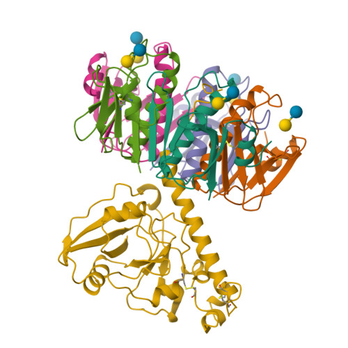
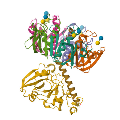
Monoclinic form of Trypanosoma cruzi trans-sialidase, in complex with 3-deoxy-2,3-dehydro-N-acetylneuraminic acid (DANA)and lactose

Buschiazzo, A., Amaya, M.F., Cremona, M.L., Frasch, A.C., Alzari, P.M.

(2002) Mol Cell 10: 757-768

Released2003-03-25
Method
X-RAY DIFFRACTION 2.5 Å
Organisms
Trypanosoma cruzi
Macromolecule
trans-sialidase (protein)
Unique LigandsDAN
Unique branched monosaccharidesBGC, GAL
Gallery item
 Explore in 3D

1MS9 | pdb_00001ms9

Download FileView File

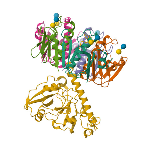
Triclinic form of Trypanosoma cruzi trans-sialidase, in complex with lactose

Buschiazzo, A., Amaya, M.F., Cremona, M.L., Frasch, A.C., Alzari, P.M.

(2002) Mol Cell 10: 757-768

Released2003-03-25
Method
X-RAY DIFFRACTION 1.58 Å
Organisms
Trypanosoma cruzi
Macromolecule
trans-sialidase (protein)
Unique branched monosaccharidesBGC, GAL
Gallery item
 Explore in 3D

1O7O | pdb_00001o7o

Download FileView File

Roles of Individual Residues of Alpha-1,3 Galactosyltransferases in Substrate Binding and Catalysis

Zhang, Y., Swaminathan, G.J., Deshpande, A., Natesh, R., Xie, Z., Acharya, K.R., Brew, K.

(2003) Biochemistry 42: 13512-13521

Released2003-11-06
Method
X-RAY DIFFRACTION 1.97 Å
Organisms
Bos taurus
Macromolecule
N-ACETYLLACTOSAMINIDE ALPHA-1,3-GALACTOSYLTRANSFERASE (protein)
Unique LigandsMN, UDP
Unique branched monosaccharidesBGC, GAL
Gallery item
 Explore in 3D

1OJJ | pdb_00001ojj

Download FileView File

Anatomy of glycosynthesis: Structure and kinetics of the Humicola insolens Cel7BE197A and E197S glycosynthase mutants

Ducros, V.M.-A., Tarling, C.A., Zechel, D.L., Brzozowski, A.M., Frandsen, T.P., Von Ossowski, I., Schulein, M., Withers, S.G., Davies, G.J.

(2003) Chem Biol 10: 619

Released2004-01-07
Method
X-RAY DIFFRACTION 1.4 Å
Organisms
Mycothermus thermophilus
Macromolecule
ENDOGLUCANASE I (protein)
Unique LigandsNAG
Unique branched monosaccharidesBGC, GAL, GLC
Gallery item
 Explore in 3D

1PUU | pdb_00001puu

Download FileView File

Mistletoe lectin I in complex with lactose

Krauspenhaar, R., Voelter, W., Stoeva, S., Mikhailov, A., Konareva, N., Betzel, C.

(2005) Acta Crystallogr Sect F Struct Biol Cryst Commun 61: 17-25

Released2004-06-25
Method
X-RAY DIFFRACTION 2.3 Å
Organisms
Viscum album
Macromolecule
lectin I A chain (protein)
lectin I B chain (protein)
Unique LigandsCL, DIO, GOL, NAG, SO4
Unique branched monosaccharidesBGC, FUL, GAL, NAG
1 to 25 of 146 Structures
Page 1 of 6
Sort by
  • About
  • About Us
  • Citing Us
  • Publications
  • Team
  • Careers
  • Usage & Privacy
  • Support
  • Contact Us
  • Help
  • Website FAQ
  • Glossary
  • Service Status
  • RCSB PDB is hosted by

  • The Rutgers Artificial Intelligence and Data Science Collaboratory logo
    Uiversity of California San Diego logoSan Diego Supercomputer Center logo
    University of California San Francisco Logo
  • RCSB PDB is a member of

  • wwPDB
    EMDataResource

  • RCSB Partners
  • Nucleic Acid Knowledgebase
  • wwPDB Partners
  • RCSB PDB
  • PDBe
  • PDBj
  • BMRB
  • EMDB
CoreTrustSeal
Global Biodata Coalition Logo

RCSB PDB Core Operations are funded by the U.S. National Science Foundation (DBI-2321666), the US Department of Energy (DE-SC0019749), and the National Cancer Institute, National Institute of Allergy and Infectious Diseases, and National Institute of General Medical Sciences of the National Institutes of Health under grant R01GM157729.