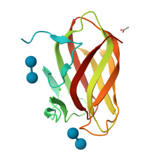
Warning You are using a web browser that we do not support. Our website will not work properly. Please update to a newer version or download a new web browser, such as Chrome or Firefox.
Help
Contact us
  • Sign in with Google
  • Sign in with Facebook
  • Sign in with ORCID

  • Help
RCSB PDB
  • Deposit 

      Prepare Data

    • PDBx/mmCIF file
    • pdb_extract
    • SF-Tool
    • Ligand Expo
    • MAXIT

      Validate Data

    • Validation Server
    • Validation API
    • Information for Journals
    • Validation Task Forces

      Deposit Data

    • wwPDB OneDep System
    • PDB-IHM

      Help and Resources

    • Deposit FAQ
    • Validation FAQ
    • Tutorials
    • Annotation Policies
    • Processing Procedures
    • PDBx/mmCIF Dictionary
    • PDBx/mmCIF User Guide
    • Chemical Component Dictionary
    • Biologically Interesting Molecule Reference Dictionary (BIRD)
    • BioSync/Beamlines/Facilities
    • Related Tools
  • Search 
    • Advanced Search
    • Sequence Similarity Search
    • Structure Similarity Search
    • Chemical Similarity Search
    • Chemical Sketch Tool
    • Browse by Annotations
    • New Entries
    • Unreleased Entries
    • PDB Statistics
  • Visualize 
    • Mol* (MolStar)
    • Sequence Annotations Viewer
    • Genome View
  • Analyze 
    • Pairwise Structure Alignment
    • Symmetry Resources in the PDB
    • Structure Quality
    • Grouping Structures
    • PDB Citation MeSH Network Explorer
    • PDB Statistics
    • EPPIC Biological Assemblies
    • External Data and Resources
    • Integrated Resources
    • Additional Resources
  • Download 
    • Coordinates and Experimental Data
    • Sequences
    • Ligands
    • File Download Services
    • Web APIs
  • Learn 

    • Training Courses
    • Guide to PDB Data
    • Molecule of the Month
    • Educational Resources
    • Curricula
    • Browse
    • News
    • SciArt Galleries
    • Irving Geis
    • David Goodsell
  • About 
    • Contact Us
    • About RCSB PDB
    • Vision and Mission
    • Citation, Usage, Privacy Policies, Logo
    • News
    • PDB History
    • PDB50
    • User Community
    • Publications
    • RCSB PDB Advisory Committee
    • Team Members
    • Service Status
  • Careers
  • COVID-19
RCSB PDB
236,974
Structures from the PDB archive
1,068,577
Computed Structure Models (CSM)
Include CSM
Advanced Search
|
Browse Annotations
Help
  • PDB-101
  • wwPDB
  • EMDataResource
  • NAKB: Nucleic Acid Knowledgebase
  • wwPDB Foundation
  • PDB-IHM Logo
  • Search
  • Query History
  • Browse Annotations
  • MyPDB
QUERY: BIRD Name HAS EXACT PHRASE "=beta-laminaribiose"
MyPDB Login
 Search API
Advanced Search Query Builder
Help
Full Text
Structure Attributes
Chemical Attributes
Help
x
NOT
Count
Add Subquery
Sequence Similarity
Sequence Motif
Structure Similarity
Structure Motif
Chemical Similarity
Return
grouped by
Include Computed Structure Models (CSM)
Count
Clear
Search
Search Summary This query matches 43 Structures.
Refinements
Structure Determination Methodology
Scientific Name of Source Organism
More...
Taxonomy
Experimental Method
Polymer Entity Type
Refinement Resolution (Å)
Release Date
Enzyme Classification Name
Membrane Protein Annotation
Symmetry Type
SCOP Classification
1 to 25 of 43 Structures
Page 1 of 2
Sort by
Gallery item
 Explore in 3D

1OD3 | pdb_00001od3

Download FileView File

Structure of CSCBM6-3 From Clostridium stercorarium in complex with laminaribiose

Boraston, A.B., Notenboom, V., Warren, R.A.J., Kilburn, D.G., Rose, D.R., Davies, G.J.

(2003) J Mol Biology 327: 659

Released2003-03-13
Method
X-RAY DIFFRACTION 1 Å
Organisms
Thermoclostridium stercorarium
Macromolecule
PUTATIVE XYLANASE (protein)
Unique LigandsACY, CA
Unique branched monosaccharidesBGC
Gallery item
 Explore in 3D

2BN0 | pdb_00002bn0

Download FileView File

Banana Lectin bound to Laminaribiose

Meagher, J.L., Winter, H.C., Ezell, P., Goldstein, I.J., Stuckey, J.A.

(2005) Glycobiology 15: 1033

Released2005-06-16
Method
X-RAY DIFFRACTION 2.8 Å
Organisms
Musa acuminata
Macromolecule
RIPENING-ASSOCIATED PROTEIN (protein)
Unique LigandsCD, SO4
Unique branched monosaccharidesBGC
Gallery item
 Explore in 3D

2CIP | pdb_00002cip

Download FileView File

Structure of the Michaelis complex of a family 26 lichenase

Money, V.A., Smith, N.L., Scaffidi, A., Stick, R.V., Gilbert, H.J., Davies, G.J.

(2006) Angew Chem Int Ed Engl 45: 5136

Released2006-07-12
Method
X-RAY DIFFRACTION 1.4 Å
Organisms
Acetivibrio thermocellus
Macromolecule
ENDOGLUCANASE H (protein)
Unique LigandsZZ1
Unique branched monosaccharidesBGC
Gallery item
 Explore in 3D

2W39 | pdb_00002w39

Download FileView File

Glc(beta-1-3)Glc disaccharide in -1 and -2 sites of Laminarinase 16A from Phanerochaete chrysosporium

Vasur, J., Kawai, R., Andersson, E., Igarashi, K., Sandgren, M., Samejima, M., Stahlberg, J.

(2009) FEBS J 276: 3858

Released2009-07-21
Method
X-RAY DIFFRACTION 1.1 Å
Organisms
Phanerodontia chrysosporium
Macromolecule
PUTATIVE LAMINARINASE (protein)
Unique LigandsLGC, NAG
Unique branched monosaccharidesBGC
Gallery item
 Explore in 3D

3AHT | pdb_00003aht

Download FileView File

Crystal structure of rice BGlu1 E176Q mutant in complex with laminaribiose

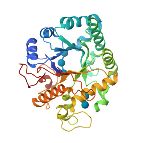
Chuenchor, W., Pengthaisong, S., Robinson, R.C., Yuvaniyama, J., Svasti, J., Ketudat Cairns, J.R.

(2011) J Struct Biol 173: 169-179

Released2010-06-02
Method
X-RAY DIFFRACTION 2.8 Å
Organisms
Oryza sativa Japonica Group
Macromolecule
Beta-glucosidase 7 (protein)
Unique LigandsMES, SO4, ZN
Unique branched monosaccharidesBGC
Gallery item
 Explore in 3D

3F5L | pdb_00003f5l

Download FileView File

Semi-active E176Q mutant of rice BGlu1, a plant exoglucanase/beta-glucosidase

Chuenchor, W., Ketudat Cairns, J.R., Pengthaisong, S., Robinson, R.C., Yuvaniyama, J., Chen, C.-J.

(2011) J Struct Biol 173: 169-179

Released2009-11-03
Method
X-RAY DIFFRACTION 1.37 Å
Organisms
Oryza sativa Japonica Group
Macromolecule
Beta-glucosidase (protein)
Unique LigandsMES, SO4, ZN
Unique branched monosaccharidesBGC
Gallery item
 Explore in 3D

3N9K | pdb_00003n9k

Download FileView File

F229A/E292S Double Mutant of Exo-beta-1,3-glucanase from Candida albicans in Complex with Laminaritriose at 1.7 A

Nakatani, Y., Cutfield, S.M., Cutfield, J.F.

(2010) FEBS J 277: 4549-4561

Released2010-09-15
Method
X-RAY DIFFRACTION 1.7 Å
Organisms
Candida albicans
Macromolecule
Glucan 1,3-beta-glucosidase (protein)
Unique LigandsCA
Unique branched monosaccharidesBGC
Gallery item
 Explore in 3D

3P5H | pdb_00003p5h

Download FileView File

Structure of the carbohydrate-recognition domain of human Langerin with Laminaritriose

Feinberg, H., Taylor, M.E., Razi, N., McBride, R., Knirel, Y.A., Graham, S.A., Drickamer, K., Weis, W.I.

(2011) J Mol Biology 405: 1027-1039

Released2010-12-08
Method
X-RAY DIFFRACTION 1.6051 Å
Organisms
Homo sapiens
Macromolecule
C-type lectin domain family 4 member K (protein)
Unique LigandsBGC, CA
Unique branched monosaccharidesBGC
Gallery item
 Explore in 3D

3VY6 | pdb_00003vy6

Download FileView File

Crystal structure of human pancreatic secretory protein ZG16p with laminaribiose

Kanagawa, M., Yamaguchi, Y.

(2014) J Biological Chem 289: 16954-16965

Released2013-09-25
Method
X-RAY DIFFRACTION 2 Å
Organisms
Homo sapiens
Macromolecule
Zymogen granule membrane protein 16 (protein)
Unique LigandsCL
Unique branched monosaccharidesBGC
Gallery item
 Explore in 3D

3WY6 | pdb_00003wy6

Download FileView File

Structure of hyperthermophilic family 12 endocellulase (E197A) from Pyrococcus furiosus in complex with laminaribiose

Kataoka, M., Ishikawa, K.

To be published

Released2015-09-16
Method
X-RAY DIFFRACTION 1.45 Å
Organisms
Pyrococcus furiosus
Macromolecule
Endoglucanase A (protein)
Unique LigandsBGC, CA, GOL, NHE
Unique branched monosaccharidesBGC
Gallery item
 Explore in 3D

4GZJ | pdb_00004gzj

Download FileView File

Active-site mutant of potato endo-1,3-beta-glucanase in complex with laminaratriose and laminaratetrose

Wojtkowiak, A., Witek, K., Hennig, J., Jaskolski, M.

(2013) Acta Crystallogr D Biol Crystallogr 69: 52-62

Released2013-01-02
Method
X-RAY DIFFRACTION 1.55 Å
Organisms
Solanum tuberosum
Macromolecule
Glucan endo-1,3-beta-D-glucosidase (protein)
Unique branched monosaccharidesBGC
Gallery item
 Explore in 3D

4JSD | pdb_00004jsd

Download FileView File

The X-ray crystal structure of a thermophilic cellobiose binding protein bound with laminaribiose

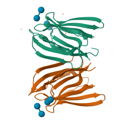
Munshi, P., Cuneo, M.J.

(2013) BMC Struct Biol 13: 18-18

Released2013-10-09
Method
X-RAY DIFFRACTION 2.05 Å
Organisms
Thermotoga maritima MSB8
Macromolecule
Oligopeptide ABC transporter, periplasmic oligopeptide-binding protein (protein)
Unique LigandsCA
Unique branched monosaccharidesBGC
Gallery item
 Explore in 3D

4R2F | pdb_00004r2f

Download FileView File

Crystal structure of sugar transporter ACHL_0255 from Arthrobacter chlorophenolicus A6, target EFI-510633, with bound laminaribiose

Patskovsky, Y., Toro, R., Bhosle, R., Al Obaidi, N., Chamala, S., Attonito, J.D., Scott Glenn, A., Chowdhury, S., Lafleur, J., Siedel, R.D., Hillerich, B., Love, J., Whalen, K.L., Gerlt, J.A., Almo, S.C., Enzyme Function Initiative (EFI)

To be published

Released2014-08-27
Method
X-RAY DIFFRACTION 2 Å
Organisms
Pseudarthrobacter chlorophenolicus A6
Macromolecule
Extracellular solute-binding protein family 1 (protein)
Unique branched monosaccharidesBGC
Gallery item
 Explore in 3D

4WTR | pdb_00004wtr

Download FileView File

Active-site mutant of Rhizomucor miehei beta-1,3-glucanosyltransferase in complex with laminaribiose

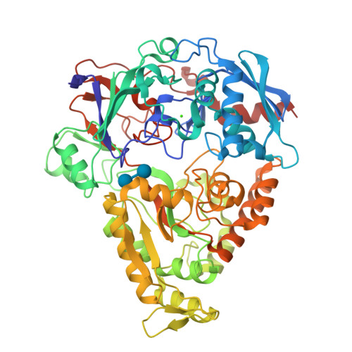
Qin, Z., Yan, Q., Lei, J., Yang, S., Jiang, Z.

(2015) Acta Crystallogr D Biol Crystallogr 71: 1714-1724

Released2015-08-12
Method
X-RAY DIFFRACTION 2.27 Å
Organisms
Rhizomucor miehei CAU432
Macromolecule
beta-1,3-glucanosyltransferase (protein)
Unique branched monosaccharidesBGC
Gallery item
 Explore in 3D

5T4G | pdb_00005t4g

Download FileView File

Crystal structure of BhGH81 in complex with laminarin

Pluvinage, B., Boraston, A.B.

(2017) Structure 25: 1348-1359.e3

Released2017-06-28
Method
X-RAY DIFFRACTION 1.8 Å
Organisms
Halalkalibacterium halodurans C-125
Macromolecule
Glycoside Hydrolase (protein)
Unique LigandsEDO, PO4
Unique branched monosaccharidesBGC, GLC
Gallery item
 Explore in 3D

5U6N | pdb_00005u6n

Download FileView File

Crystal structure of UDP-glucosyltransferase, UGT74F2 (T15S), with UDP and salicylic acid

George Thompson, A.M., Iancu, C.V., Dean, J.V., Choe, J.

(2017) Sci Rep 7: 46629-46629

Released2017-05-03
Method
X-RAY DIFFRACTION 2.001 Å
Organisms
Arabidopsis thaliana
Macromolecule
UDP-glycosyltransferase 74F2 (protein)
Unique LigandsBGC, GLN, SAL, UDP
Unique branched monosaccharidesBGC
Gallery item
 Explore in 3D

5U6S | pdb_00005u6s

Download FileView File

Crystal structure of UDP-glucosyltransferase, UGT74F2, with UDP and 2-bromobenzoic acid

George Thompson, A.M., Iancu, C.V., Dean, J.V., Choe, J.

(2017) Sci Rep 7: 46629-46629

Released2017-05-03
Method
X-RAY DIFFRACTION 1.996 Å
Organisms
Arabidopsis thaliana
Macromolecule
UDP-glycosyltransferase 74F2 (protein)
Unique Ligands7WV, BGC, UDP
Unique branched monosaccharidesBGC
Gallery item
 Explore in 3D

5UPI | pdb_00005upi

Download FileView File

Crystal structure of BhGH81 mutant in complex with laminaro-biose

Pluvinage, B., Boraston, A.B.

(2017) Structure

Released2017-06-28
Method
X-RAY DIFFRACTION 1.65 Å
Organisms
Halalkalibacterium halodurans C-125
Macromolecule
BH0236 protein (protein)
Unique LigandsEDO, PO4
Unique branched monosaccharidesBGC
Gallery item
 Explore in 3D

5UPM | pdb_00005upm

Download FileView File

Crystal structure of BhGH81 mutant in complex with laminaro-triose

Pluvinage, B., Boraston, A.B.

(2017) Structure

Released2017-06-28
Method
X-RAY DIFFRACTION 1.7 Å
Organisms
Halalkalibacterium halodurans C-125
Macromolecule
BH0236 protein (protein)
Unique LigandsEDO, PO4
Unique branched monosaccharidesBGC, GLC
Gallery item
 Explore in 3D

5UPN | pdb_00005upn

Download FileView File

Crystal structure of BhGH81 mutant in complex with laminaro-tetraose

Pluvinage, B., Boraston, A.B.

(2017) Structure

Released2017-06-28
Method
X-RAY DIFFRACTION 1.8 Å
Organisms
Halalkalibacterium halodurans C-125
Macromolecule
BH0236 protein (protein)
Unique LigandsBGC, EDO, GLC, PO4
Unique branched monosaccharidesBGC
Gallery item
 Explore in 3D

5V1W | pdb_00005v1w

Download FileView File

Crystal structure of BhGH81 in complex with laminaro-biose

Pluvinage, B., Boraston, A.B.

(2017) Structure

Released2017-06-28
Method
X-RAY DIFFRACTION 2.15 Å
Organisms
Halalkalibacterium halodurans C-125
Macromolecule
Glycoside Hydrolase (protein)
Unique LigandsEDO, PO4
Unique branched monosaccharidesBGC
Gallery item
 Explore in 3D

5V2J | pdb_00005v2j

Download FileView File

Crystal structure of UDP-glucosyltransferase, UGT74F2 (T15S), with UDP and 2-bromobenzoic acid

George Thompson, A.M., Iancu, C.V., Dean, J.V., Choe, J.

(2017) Sci Rep 7: 46629-46629

Released2017-05-03
Method
X-RAY DIFFRACTION 1.8 Å
Organisms
Arabidopsis thaliana
Macromolecule
UDP-glycosyltransferase 74F2 (protein)
Unique Ligands7WV, BGC, UDP
Unique branched monosaccharidesBGC
Gallery item
 Explore in 3D

5V2K | pdb_00005v2k

Download FileView File

Crystal structure of UDP-glucosyltransferase, UGT74F2 (T15A), with UDP and 2-bromobenzoic acid

George Thompson, A.M., Iancu, C.V., Dean, J.V., Choe, J.

(2017) Sci Rep 7: 46629-46629

Released2017-05-03
Method
X-RAY DIFFRACTION 2.002 Å
Organisms
Arabidopsis thaliana
Macromolecule
UDP-glycosyltransferase 74F2 (protein)
Unique Ligands7WV, BGC, UDP
Unique branched monosaccharidesBGC
Gallery item
 Explore in 3D

5XC2 | pdb_00005xc2

Download FileView File

Crystal structure of GH family 81 beta-1,3-glucanase from Rhizomucr miehei complexed with laminarihexaose

Qin, Z., Yang, S., Peng, Z., Yan, Q., Jiang, Z.

To be published

Released2018-03-28
Method
X-RAY DIFFRACTION 2.7 Å
Organisms
Rhizomucor miehei
Macromolecule
Endo-beta-1,3-glucanase (protein)
Unique branched monosaccharidesBGC
Gallery item
 Explore in 3D

6EV2 | pdb_00006ev2

Download FileView File

Crystal structure of antibody against schizophyllan in complex with laminarihexaose

Sung, K.H., Josewski, J., Duebel, S., Blankenfeldt, W., Rau, U.

(2018) Sci Rep 8: 13652-13652

Released2018-09-26
Method
X-RAY DIFFRACTION 2.403 Å
Organisms
Mus musculus
Macromolecule
Heavy chain (protein)
Light chain (protein)
Unique LigandsBGC
Unique branched monosaccharidesBGC
1 to 25 of 43 Structures
Page 1 of 2
Sort by
  • About
  • About Us
  • Citing Us
  • Publications
  • Team
  • Careers
  • Usage & Privacy
  • Support
  • Contact Us
  • Help
  • Website FAQ
  • Glossary
  • Service Status
  • RCSB PDB is hosted by

  • The Rutgers Artificial Intelligence and Data Science Collaboratory logo
    Uiversity of California San Diego logoSan Diego Supercomputer Center logo
    University of California San Francisco Logo
  • RCSB PDB is a member of the

  • wwPDB
    EMDataResource

  • RCSB Partners
  • Nucleic Acid Knowledgebase
  • wwPDB Partners
  • RCSB PDB
  • PDBe
  • PDBj
  • BMRB
  • EMDB
CoreTrustSeal
Global Biodata Coalition Logo

RCSB PDB Core Operations are funded by the U.S. National Science Foundation (DBI-2321666), the US Department of Energy (DE-SC0019749), and the National Cancer Institute, National Institute of Allergy and Infectious Diseases, and National Institute of General Medical Sciences of the National Institutes of Health under grant R01GM157729.