Warning You are using a web browser that we do not support. Our website will not work properly. Please update to a newer version or download a new web browser, such as Chrome or Firefox.
Help
Contact us
  • Sign in with Google
  • Sign in with Facebook
  • Sign in with ORCID

  • Help
RCSB PDB
  • Deposit 

      Prepare Data

    • PDBx/mmCIF file
    • pdb_extract
    • SF-Tool
    • Ligand Expo
    • MAXIT

      Validate Data

    • Validation Server
    • Validation API
    • Information for Journals
    • Validation Task Forces

      Deposit Data

    • wwPDB OneDep System
    • PDB-IHM

      Help and Resources

    • Deposit FAQ
    • Validation FAQ
    • Tutorials
    • Annotation Policies
    • Processing Procedures
    • PDBx/mmCIF Dictionary
    • PDBx/mmCIF User Guide
    • Chemical Component Dictionary
    • Biologically Interesting Molecule Reference Dictionary (BIRD)
    • BioSync/Beamlines/Facilities
    • Related Tools
  • Search 
    • Advanced Search
    • Sequence Similarity Search
    • Structure Similarity Search
    • Chemical Similarity Search
    • Chemical Sketch Tool
    • Browse by Annotations
    • New Entries
    • Unreleased Entries
    • PDB Statistics
  • Visualize 
    • Mol* (MolStar)
    • Sequence Annotations Viewer
    • Genome View
  • Analyze 
    • Pairwise Structure Alignment
    • Symmetry Resources in the PDB
    • Structure Quality
    • Grouping Structures
    • PDB Citation MeSH Network Explorer
    • PDB Statistics
    • EPPIC Biological Assemblies
    • External Data and Resources
    • Integrated Resources
    • Additional Resources
  • Download 
    • Coordinates and Experimental Data
    • Sequences
    • Ligands
    • File Download Services
    • Web APIs
  • Learn 

    • Training Courses
    • Guide to PDB Data
    • Molecule of the Month
    • Educational Resources
    • Curricula
    • Browse
    • News
    • SciArt Galleries
    • Irving Geis
    • David Goodsell
  • About 
    • Contact Us
    • About RCSB PDB
    • Vision and Mission
    • Citation, Usage, Privacy Policies, Logo
    • News
    • PDB History
    • PDB50
    • User Community
    • Publications
    • RCSB PDB Advisory Committee
    • Team Members
    • Service Status
  • Careers
  • COVID-19
RCSB PDB
239,267
Structures from the PDB archive
1,068,577
Computed Structure Models (CSM)
Include CSM
Advanced Search
|
Browse Annotations
Help
  • PDB-101
  • wwPDB
  • EMDataResource
  • NAKB: Nucleic Acid Knowledgebase
  • wwPDB Foundation
  • PDB-IHM Logo
  • Search
  • Query History
  • Browse Annotations
  • MyPDB
QUERY: BIRD Type = "Glycopeptide"
MyPDB Login
 Search API
Advanced Search Query Builder
Help
Full Text
Structure Attributes
Chemical Attributes
Help
x
NOT
Count
Add Subquery
Sequence Similarity
Sequence Motif
Structure Similarity
Structure Motif
Chemical Similarity
Return
grouped by
Include Computed Structure Models (CSM)
Count
Clear
Search
Search Summary This query matches 47 Structures.
Refinements
Structure Determination Methodology
Scientific Name of Source Organism
More...
Taxonomy
Experimental Method
Polymer Entity Type
Refinement Resolution (Å)
Release Date
Enzyme Classification Name
Membrane Protein Annotation
Symmetry Type
SCOP Classification
1 to 25 of 47 Structures
Page 1 of 2
Sort by
Gallery item
 Explore in 3D

1AA5 | pdb_00001aa5

Download FileView File

VANCOMYCIN

Loll, P.J., Bevivino, A.E., Korty, B.D., Axelsen, P.H.

(1997) J Am Chem Soc 119: 1516

Released1997-08-20
Method
X-RAY DIFFRACTION 0.89 Å
Organisms
Amycolatopsis orientalis
Macromolecule
VANCOMYCIN (protein)
Unique LigandsACY, CL
Unique branched monosaccharidesBGC, RER
Gallery item
 Explore in 3D

1C0Q | pdb_00001c0q

Download FileView File

COMPLEX OF VANCOMYCIN WITH 2-ACETOXY-D-PROPANOIC ACID

Loll, P.J., Kaplan, J., Selinsky, B., Axelsen, P.H.

(1999) J Med Chem 42: 4714-4719

Released1999-07-30
Method
X-RAY DIFFRACTION 1 Å
Organisms
Amycolatopsis orientalis
Macromolecule
VANCOMYCIN (protein)
Unique LigandsCL, LAC
Unique branched monosaccharidesBGC, RER
Gallery item
 Explore in 3D

1C0R | pdb_00001c0r

Download FileView File

COMPLEX OF VANCOMYCIN WITH D-LACTIC ACID

Loll, P.J., Kaplan, J., Selinsky, B., Axelsen, P.H.

(1999) J Med Chem 42: 4714

Released1999-07-30
Method
X-RAY DIFFRACTION 1 Å
Organisms
Amycolatopsis orientalis
Macromolecule
VANCOMYCIN (protein)
Unique LigandsCL, LAC
Unique branched monosaccharidesBGC, RER
Gallery item
 Explore in 3D

1DMT | pdb_00001dmt

Download FileView File

STRUCTURE OF HUMAN NEUTRAL ENDOPEPTIDASE COMPLEXED WITH PHOSPHORAMIDON

Oefner, C., D'Arcy, A., Hennig, M., Winkler, F.K., Dale, G.E.

(2000) J Mol Biology 296: 341-349

Released2000-12-20
Method
X-RAY DIFFRACTION 2.1 Å
Organisms
Homo sapiens
Macromolecule
NEUTRAL ENDOPEPTIDASE (protein)
Unique LigandsGOL, NAG, RDF, ZN
Gallery item
 Explore in 3D

1FVM | pdb_00001fvm

Download FileView File

Complex of vancomycin with DI-acetyl-LYS-D-ALA-D-ALA

Nitanai, Y., Kakoi, K., Aoki, K.

(2009) J Mol Biology 385: 1422

Released2000-11-01
Method
X-RAY DIFFRACTION 1.8 Å
Organisms
Amycolatopsis orientalis
Macromolecule
DI-ACETYL-LYS-D-ALA-D-ALA (protein)
VANCOMYCIN (protein)
Unique branched monosaccharidesBGC, RER
Gallery item
 Explore in 3D

1GAC | pdb_00001gac

Download FileView File

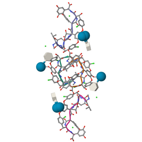
NMR structure of asymmetric homodimer of a82846b, a glycopeptide antibiotic, complexed with its cell wall pentapeptide fragment

Kline, A.D., Prowse, W.G., Skelton, M.A., Loncharich, R.J.

(1995) Biochemistry 34: 9632

Released1996-08-17
Method
SOLUTION NMR
Organisms
Amycolatopsis orientalis
Macromolecule
CELL WALL PENTAPEPTIDE (protein)
CHLOROORIENTICIN A (protein)
Unique LigandsRER
Unique branched monosaccharidesBGC, RER
Gallery item
 Explore in 3D

1GO6 | pdb_00001go6

Download FileView File

Balhimycin in complex with Lys-D-ala-D-ala

Lehmann, C., Bunkoczi, G., Vertesy, L., Sheldrick, G.M.

(2002) J Mol Biology 318: 723

Released2002-06-13
Method
X-RAY DIFFRACTION 0.98 Å
Organisms
Amycolatopsis sp.
synthetic construct
Macromolecule
BALHIMYCIN (protein)
PEPTIDE LYS-DAL-DAL (protein)
Unique LigandsBGC, CIT, DVC, MPD, MRD
Gallery item
 Explore in 3D

1HH3 | pdb_00001hh3

Download FileView File

Decaplanin first P21-Form

Lehmann, C., Vertessy, L., Sheldrick, G.M., Dauter, Z., Dauter, M.

(2003) Helvetica Chim Acta 86: 1478

Released2005-07-11
Method
X-RAY DIFFRACTION 1 Å
Organisms
uncultured actinomycete
Macromolecule
DECAPLANIN (protein)
Unique LigandsERE, GOL
Unique branched monosaccharidesBGC, RAM
Gallery item
 Explore in 3D

1HHA | pdb_00001hha

Download FileView File

Decaplanin first P6122-Form

Lehmann, C., Vertessy, L., Sheldrick, G.M., Dauter, Z., Dauter, M.

(2003) Helvetica Chim Acta 86: 1478

Released2005-07-11
Method
X-RAY DIFFRACTION 1.9 Å
Organisms
uncultured actinomycete
Macromolecule
DECAPLANIN (protein)
Unique LigandsERE, GOL
Unique branched monosaccharidesBGC, RAM
Gallery item
 Explore in 3D

1HHC | pdb_00001hhc

Download FileView File

Crystal structure of Decaplanin - space group P21, second form

Lehmann, C., Vertessy, L., Sheldrick, G.M., Dauter, Z., Dauter, M.

(2003) Helvetica Chim Acta 86: 1478

Released2005-07-11
Method
X-RAY DIFFRACTION 1.13 Å
Organisms
uncultured actinomycete
Macromolecule
DECAPLANIN (protein)
Unique LigandsCIT, ERE
Unique branched monosaccharidesBGC, RAM
Gallery item
 Explore in 3D

1HHF | pdb_00001hhf

Download FileView File

Decaplanin second P6122-Form

Lehmann, C., Vertessy, L., Sheldrick, G.M., Dauter, Z., Dauter, M.

(2003) Helvetica Chim Acta 86: 1478

Released2005-07-11
Method
X-RAY DIFFRACTION 1.47 Å
Organisms
uncultured actinomycete
Macromolecule
DECAPLANIN (protein)
Unique LigandsCL, ERE, PO4
Unique branched monosaccharidesBGC, RAM
Gallery item
 Explore in 3D

1HHU | pdb_00001hhu

Download FileView File

Balhimycin in complex with D-Ala-D-Ala

Lehmann, C., Bunkoczi, G., Sheldrick, G.M., Vertessy, L.

(2002) J Mol Biology 318: 723

Released2003-09-05
Method
X-RAY DIFFRACTION 0.89 Å
Organisms
Amycolatopsis sp.
Macromolecule
BALHIMYCIN (protein)
Unique LigandsBGC, CIT, DAL, DVC, MPD
Gallery item
 Explore in 3D

1HHY | pdb_00001hhy

Download FileView File

Deglucobalhimycin in complex with D-Ala-D-Ala

Lehmann, C., Bunkoczi, G., Sheldrick, G.M., Vertesy, L.

(2002) J Mol Biology 318: 723

Released2003-09-05
Method
X-RAY DIFFRACTION 0.89 Å
Organisms
Amycolatopsis sp.
Macromolecule
DEGLUCOBALHIMYCIN (protein)
Unique LigandsDAL, DVC, NA, PGO
Gallery item
 Explore in 3D

1HHZ | pdb_00001hhz

Download FileView File

Deglucobalhimycin in complex with cell wall pentapeptide

Lehmann, C., Bunkoczi, G., Sheldrick, G.M., Vertesy, L.

(2002) J Mol Biology 318: 723

Released2003-09-05
Method
X-RAY DIFFRACTION 0.99 Å
Organisms
Amycolatopsis sp.
synthetic construct
Macromolecule
CELL WALL PEPTIDE (protein)
DEGLUCOBALHIMYCIN (protein)
Unique LigandsDVC, PGR
Gallery item
 Explore in 3D

1PN3 | pdb_00001pn3

Download FileView File

Crystal Structure of TDP-epi-Vancosaminyltransferase GtfA in complexes with TDP and the acceptor substrate DVV.

Mulichak, A.M., Losey, H.C., Lu, W., Wawrzak, Z., Walsh, C.T., Garavito, R.M.

(2003) Proc Natl Acad Sci U S A 100: 9238

Released2003-08-12
Method
X-RAY DIFFRACTION 2.8 Å
Organisms
Amycolatopsis orientalis
Macromolecule
DESVANCOSAMINYL VANCOMYCIN (protein)
GLYCOSYLTRANSFERASE GTFA (protein)
Unique LigandsBGC, TYD
Gallery item
 Explore in 3D

1PNV | pdb_00001pnv

Download FileView File

Crystal Structure of TDP-epi-Vancosaminyltransferase GtfA in complexes with TDP and Vancomycin

Mulichak, A.M., Losey, H.C., Lu, W., Wawrzak, Z., Walsh, C.T., Garavito, R.M.

(2003) Proc Natl Acad Sci U S A 100: 9238

Released2003-08-12
Method
X-RAY DIFFRACTION 2.8 Å
Organisms
Amycolatopsis orientalis
Macromolecule
GLYCOSYLTRANSFERASE GTFA (protein)
VANCOMYCIN (protein)
Unique LigandsTYD
Unique branched monosaccharidesBGC, RER
Gallery item
 Explore in 3D

1QD8 | pdb_00001qd8

Download FileView File

COMPLEX OF VANCOMYCIN WITH N-ACETYL GLYCINE

Loll, P.J., Kaplan, J., Selinsky, B., Axelsen, P.H.

(1999) J Med Chem 42: 4714

Released1999-08-16
Method
X-RAY DIFFRACTION 1 Å
Organisms
Amycolatopsis orientalis
Macromolecule
VANCOMYCIN (protein)
Unique LigandsAAC, CL
Unique branched monosaccharidesBGC, RER
Gallery item
 Explore in 3D

1RRV | pdb_00001rrv

Download FileView File

X-ray crystal structure of TDP-vancosaminyltransferase GtfD as a complex with TDP and the natural substrate, desvancosaminyl vancomycin.

Mulichak, A.M., Lu, W., Losey, H.C., Walsh, C.T., Garavito, R.M.

(2004) Biochemistry 43: 5170

Released2004-05-18
Method
X-RAY DIFFRACTION 2 Å
Organisms
Amycolatopsis orientalis
Macromolecule
DESVANCOSAMINYL VANCOMYCIN (protein)
GLYCOSYLTRANSFERASE GTFD (protein)
Unique LigandsBGC, GOL, K, TYD
Gallery item
 Explore in 3D

1SHO | pdb_00001sho

Download FileView File

CRYSTAL STRUCTURE OF VANCOMYCIN AT ATOMIC RESOLUTION

Sheldrick, G.M.

(1996) Structure 4: 1509

Released1997-12-24
Method
X-RAY DIFFRACTION 1.09 Å
Organisms
Amycolatopsis orientalis
Macromolecule
VANCOMYCIN (protein)
Unique LigandsACT, CL
Unique branched monosaccharidesBGC, RER
Gallery item
 Explore in 3D

1TLP | pdb_00001tlp

Download FileView File

CRYSTALLOGRAPHIC STRUCTURAL ANALYSIS OF PHOSPHORAMIDATES AS INHIBITORS AND TRANSITION-STATE ANALOGS OF THERMOLYSIN

Tronrud, D.E., Monzingo, A.F., Matthews, B.W.

(1986) Eur J Biochem 157: 261-268

Released1989-01-09
Method
X-RAY DIFFRACTION 2.3 Å
Organisms
Bacillus thermoproteolyticus
Macromolecule
THERMOLYSIN (protein)
Unique LigandsCA, RDF, ZN
Gallery item
 Explore in 3D

2XAD | pdb_00002xad

Download FileView File

Crystal structure of deacetylase-teicoplanin complex in biosynthesis pathway of teicoplanin

Chan, H.C., Huang, Y.T., Lyu, S.Y., Huang, C.J., Li, Y.S., Liu, Y.C., Chou, C.C., Tsai, M.D., Li, T.L.

(2011) Mol Biosyst 7: 1224

Released2011-03-02
Method
X-RAY DIFFRACTION 1.7 Å
Organisms
Actinoplanes teichomyceticus
Nonomuraea gerenzanensis
Macromolecule
N-ACYL GLM PEUDO-TEICOPLANIN DEACETYLASE (protein)
TEICOPLANIN (protein)
Unique LigandsBMA, GCS, NAG, T55
Gallery item
 Explore in 3D

3DBK | pdb_00003dbk

Download FileView File

Pseudomonas aeruginosa elastase with phosphoramidon

McKay, D.B., Overgaard, M.T.

To be published

Released2009-07-07
Method
X-RAY DIFFRACTION 1.4 Å
Organisms
Pseudomonas aeruginosa
Macromolecule
Elastase (protein)
Unique LigandsCA, RDF, SO4, ZN
Gallery item
 Explore in 3D

3DWB | pdb_00003dwb

Download FileView File

structure of human ECE-1 complexed with phosphoramidon

Oefner, C.

(2009) J Mol Biology 385: 178-187

Released2008-11-25
Method
X-RAY DIFFRACTION 2.38 Å
Organisms
Homo sapiens
Macromolecule
Endothelin-converting enzyme 1 (protein)
Unique Ligands5HD, RDF, ZN
Gallery item
 Explore in 3D

3RUN | pdb_00003run

Download FileView File

New strategy to analyze structures of glycopeptide antibiotic-target complexes

Economou, N.J., Townsend, T.M., Loll, P.J.

(2012) J Am Chem Soc 134: 4637-4645

Released2012-06-06
Method
X-RAY DIFFRACTION 1.4 Å
Organisms
Amycolatopsis orientalis
Tequatrovirus T4
Macromolecule
LYSOZYME (protein)
VANCOMYCIN (protein)
Unique LigandsCL, IPA, MPD, MRD, PO4, TRS
Unique branched monosaccharidesBGC, RER
Gallery item
 Explore in 3D

3VFJ | pdb_00003vfj

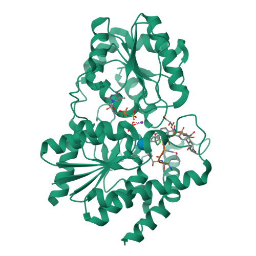
Download FileView File

The structure of monodechloro-teicoplanin in complex with its ligand, using MBP as a ligand carrier

Economou, N.J., Weeks, S.D., Grasty, K.C., Loll, P.J.

(2013) Acta Crystallogr D Biol Crystallogr 69: 520-533

Released2013-01-09
Method
X-RAY DIFFRACTION 2.05 Å
Organisms
Actinoplanes teichomyceticus
Escherichia coli K-12
Macromolecule
Maltose-binding periplasmic protein, C-terminal fused by Cys-Lys-D-Ala-D-Ala (protein)
MonodeChloro- Teicoplanin A2-2 (protein)
Unique LigandsACT, CAC, GCS, MAN, NAG, T55, ZN
1 to 25 of 47 Structures
Page 1 of 2
Sort by
  • About
  • About Us
  • Citing Us
  • Publications
  • Team
  • Careers
  • Usage & Privacy
  • Support
  • Contact Us
  • Help
  • Website FAQ
  • Glossary
  • Service Status
  • RCSB PDB is hosted by

  • The Rutgers Artificial Intelligence and Data Science Collaboratory logo
    Uiversity of California San Diego logoSan Diego Supercomputer Center logo
    University of California San Francisco Logo
  • RCSB PDB is a member of the

  • wwPDB
    EMDataResource

  • RCSB Partners
  • Nucleic Acid Knowledgebase
  • wwPDB Partners
  • RCSB PDB
  • PDBe
  • PDBj
  • BMRB
  • EMDB
CoreTrustSeal
Global Biodata Coalition Logo

RCSB PDB Core Operations are funded by the U.S. National Science Foundation (DBI-2321666), the US Department of Energy (DE-SC0019749), and the National Cancer Institute, National Institute of Allergy and Infectious Diseases, and National Institute of General Medical Sciences of the National Institutes of Health under grant R01GM157729.