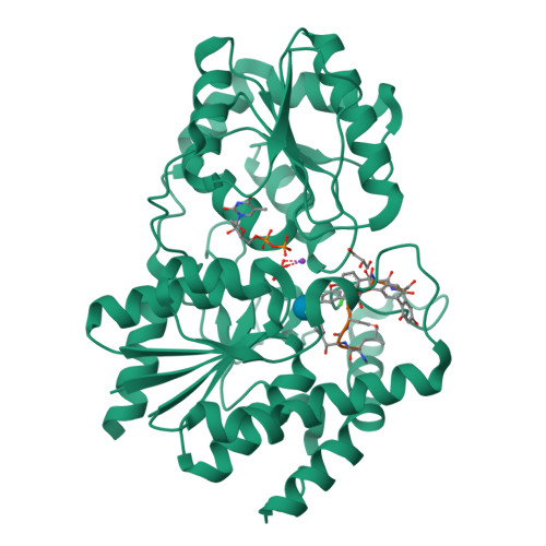
1RRV

X-ray crystal structure of TDP-vancosaminyltransferase GtfD as a complex with TDP and the natural substrate, desvancosaminyl vancomycin.


Experimental Data Snapshot

  • Method: X-RAY DIFFRACTION
  • Resolution: 2.00 Å
  • R-Value Free: 0.252 
  • R-Value Work: 0.210 
  • R-Value Observed: 0.210 

Starting Model: experimental
View more details

wwPDB Validation   3D Report Full Report


Ligand Structure Quality Assessment 


This is version 2.2 of the entry. See complete history


Literature

Crystal Structure of Vancosaminyltransferase Gtfd from the Vancomycin Biosynthetic Pathway: Interactions with Acceptor and Nucleotide Ligands

Mulichak, A.M.Lu, W.Losey, H.C.Walsh, C.T.Garavito, R.M.

(2004) Biochemistry 43: 5170

  • DOI: https://doi.org/10.1021/bi036130c
  • Primary Citation of Related Structures:  
    1RRV

  • PubMed Abstract: 

    The TDP-vancosaminyltransferase GtfD catalyzes the attachment of L-vancosamine to a monoglucosylated heptapeptide intermediate during the final stage of vancomycin biosynthesis. Glycosyltransferases from this and similar antibiotic pathways are potential tools for the design of new compounds that are effective against vancomycin resistant bacterial strains. We have determined the X-ray crystal structure of GtfD as a complex with TDP and the natural glycopeptide substrate at 2.0 A resolution. GtfD, a member of the bidomain GT-B glycosyltransferase superfamily, binds TDP in the interdomain cleft, while the aglycone acceptor binds in a deep crevice in the N-terminal domain. However, the two domains are more interdependent in terms of substrate binding and overall structure than was evident in the structures of closely related glycosyltransferases GtfA and GtfB. Structural and kinetic analyses support the identification of Asp13 as a catalytic general base, with a possible secondary role for Thr10. Several residues have also been identified as being involved in donor sugar binding and recognition.


  • Organizational Affiliation

    Department of Biochemistry and Molecular Biology, Michigan State University, East Lansing, Michigan 48824-1319, USA.


Macromolecules
Find similar proteins by:  (by identity cutoff)  |  3D Structure
Entity ID: 1
MoleculeChains Sequence LengthOrganismDetailsImage
GLYCOSYLTRANSFERASE GTFD
A, B
416Amycolatopsis orientalisMutation(s): 0 
Gene Names: GTFD
EC: 2.4.1.322
UniProt
Find proteins for Q9AFC7 (Amycolatopsis orientalis)
Explore Q9AFC7 
Go to UniProtKB:  Q9AFC7
Entity Groups  
Sequence Clusters30% Identity50% Identity70% Identity90% Identity95% Identity100% Identity
UniProt GroupQ9AFC7
Sequence Annotations
Expand
  • Reference Sequence

Find similar proteins by:  Sequence   |   3D Structure  

Entity ID: 2
MoleculeChains Sequence LengthOrganismDetailsImage
DESVANCOSAMINYL VANCOMYCIN
C, D
7Amycolatopsis orientalisMutation(s): 0 
Sequence Annotations
Expand
  • Reference Sequence
Small Molecules
Ligands 4 Unique
IDChains Name / Formula / InChI Key2D Diagram3D Interactions
TYD
Query on TYD

Download Ideal Coordinates CCD File 
F [auth A],
J [auth B]
THYMIDINE-5'-DIPHOSPHATE
C10 H16 N2 O11 P2
UJLXYODCHAELLY-XLPZGREQSA-N
BGC
Query on BGC

Download Ideal Coordinates CCD File 
L [auth C],
M [auth D]
beta-D-glucopyranose
C6 H12 O6
WQZGKKKJIJFFOK-VFUOTHLCSA-N
GOL
Query on GOL

Download Ideal Coordinates CCD File 
G [auth A],
H [auth A],
K [auth B]
GLYCEROL
C3 H8 O3
PEDCQBHIVMGVHV-UHFFFAOYSA-N
K
Query on K

Download Ideal Coordinates CCD File 
E [auth A],
I [auth B]
POTASSIUM ION
K
NPYPAHLBTDXSSS-UHFFFAOYSA-N
Modified Residues  1 Unique
IDChains TypeFormula2D DiagramParent
OMY
Query on OMY
C, D
L-PEPTIDE LINKINGC9 H10 Cl N O4TYR
Biologically Interesting Molecules (External Reference) 1 Unique
Experimental Data & Validation

Experimental Data

  • Method: X-RAY DIFFRACTION
  • Resolution: 2.00 Å
  • R-Value Free: 0.252 
  • R-Value Work: 0.210 
  • R-Value Observed: 0.210 
  • Space Group: P 1 21 1
Unit Cell:
Length ( Å )Angle ( ˚ )
a = 50.67α = 90
b = 64.12β = 91.73
c = 144.08γ = 90
Software Package:
Software NamePurpose
AMoREphasing
CNSrefinement
MAR345data collection
XDSdata scaling

Structure Validation

View Full Validation Report


Ligand Structure Quality Assessment 


Entry History 

Deposition Data

Revision History  (Full details and data files)

  • Version 1.0: 2004-05-18
    Type: Initial release
  • Version 1.1: 2011-06-14
    Changes: Version format compliance
  • Version 1.2: 2011-07-13
    Changes: Version format compliance
  • Version 1.3: 2011-07-27
    Changes: Atomic model, Database references, Derived calculations, Non-polymer description, Structure summary
  • Version 1.4: 2012-12-12
    Changes: Other
  • Version 1.5: 2017-10-11
    Changes: Refinement description
  • Version 1.6: 2017-11-01
    Changes: Derived calculations
  • Version 2.0: 2020-07-29
    Type: Remediation
    Reason: Carbohydrate remediation
    Changes: Data collection, Database references, Derived calculations, Polymer sequence, Structure summary
  • Version 2.1: 2023-08-23
    Changes: Data collection, Database references, Refinement description, Structure summary
  • Version 2.2: 2023-11-15
    Changes: Data collection, Derived calculations