


Full Text |
QUERY: Source Organism Taxonomy Name (Full Lineage) = "Clostridium cochlearium" | MyPDB Login | Search API |
| Search Summary | This query matches 6 Structures. |
Structure Determination MethodologyScientific Name of Source OrganismTaxonomyExperimental MethodPolymer Entity TypeRefinement Resolution (Å)Release DateEnzyme Classification NameSymmetry TypeSCOP Classification | 1 to 6 of 6 Structures Page 1 of 1 Sort by 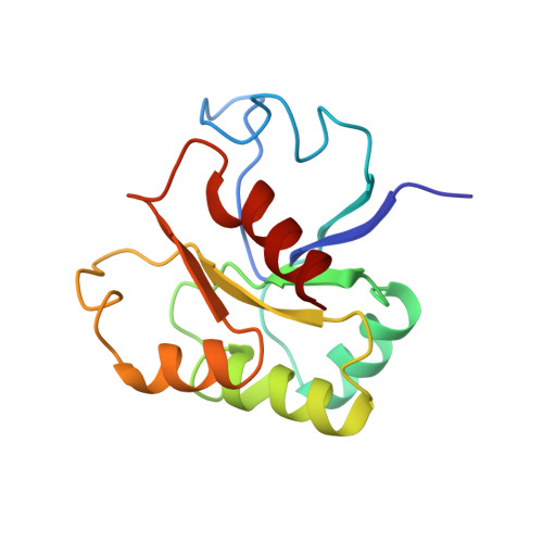
GLUTAMATE MUTASE (B12-BINDING SUBUNIT), NMR, MINIMIZED AVERAGE STRUCTUREHoffmann, B., Konrat, R., Bothe, H., Buckel, W., Kraeutler, B. (1999) Eur J Biochem 263: 178-188

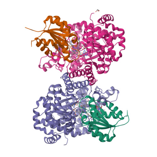
GLUTAMATE MUTASE FROM CLOSTRIDIUM COCHLEARIUM RECONSTITUTED WITH METHYL-COBALAMINGruber, K., Reitzer, R., Kratky, C. (1999) Structure 7: 891-902

STRUCTURE OF THE COENZYME B12 DEPENDENT ENZYME GLUTAMATE MUTASE FROM CLOSTRIDIUM COCHLEARIUMReitzer, R., Gruber, K., Kratky, C. (1999) Structure 7: 891-902

GLUTAMATE MUTASE FROM CLOSTRIDIUM COCHLEARIUM: COMPLEX WITH ADENOSYLCOBALAMIN AND SUBSTRATE(2001) Angew Chem Int Ed Engl 40: 3377-3380
Structure of glutamate mutase reconstituted with homo-coenzyme B12Gruber, K., Csitkovits, V., Kratky, C. (2022) Angew Chem Int Ed Engl 61: e202208295-e202208295
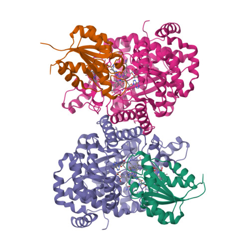
Structure of glutamate mutase reconstituted with bishomo-coenzyme B12Gruber, K., Csitkovits, V., Kratky, C. (2022) Angew Chem Int Ed Engl 61: e202208295-e202208295
1 to 6 of 6 Structures Page 1 of 1 Sort by |


