6H9F | pdb_00006h9f

Structure of glutamate mutase reconstituted with bishomo-coenzyme B12


Experimental Data Snapshot

  • Method: X-RAY DIFFRACTION
  • Resolution: 2.10 Å
  • R-Value Free: 
    0.203 (Depositor), 0.200 (DCC) 
  • R-Value Work: 
    0.158 (Depositor), 0.160 (DCC) 
  • R-Value Observed: 
    0.160 (Depositor) 

Starting Model: experimental
View more details

wwPDB Validation   3D Report Full Report


Ligand Structure Quality Assessment 

Created with Raphaël 2.3.0Worse 01 BetterLigand structure goodness of fit to experimental dataBest fitted B12Click on this verticalbar to view detailsBest fitted 8ZBClick on this verticalbar to view details

This is version 1.2 of the entry. See complete history


Literature

Structure-Based Demystification of Radical Catalysis by a Coenzyme B 12 Dependent Enzyme-Crystallographic Study of Glutamate Mutase with Cofactor Homologues.

Gruber, K.Csitkovits, V.Lyskowski, A.Kratky, C.Krautler, B.

(2022) Angew Chem Int Ed Engl 61: e202208295-e202208295

  • DOI: https://doi.org/10.1002/anie.202208295
  • Primary Citation of Related Structures:  
    6H9E, 6H9F

  • PubMed Abstract: 

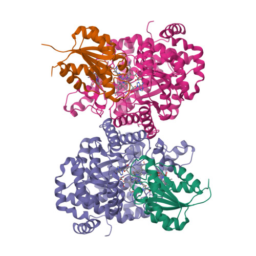
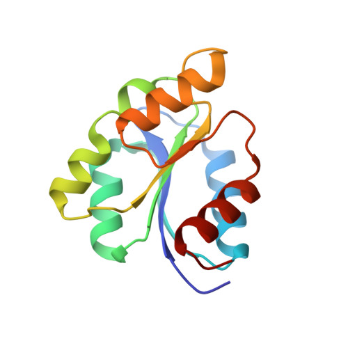
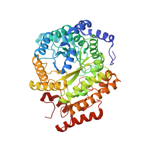
    Catalysis by radical enzymes dependent on coenzyme B 12 (AdoCbl) relies on the reactive primary 5'-deoxy-5'adenosyl radical, which originates from reversible Co-C bond homolysis of AdoCbl. This bond homolysis is accelerated roughly 10 12 -fold upon binding the enzyme substrate. The structural basis for this activation is still strikingly enigmatic. As revealed here, a displaced firm adenosine binding cavity in substrate-loaded glutamate mutase (GM) causes a structural misfit for intact AdoCbl that is relieved by the homolytic Co-C bond cleavage. Strategically interacting adjacent adenosine- and substrate-binding protein cavities provide a tight caged radical reaction space, controlling the entire radical path. The GM active site is perfectly structured for promoting radical catalysis, including "negative catalysis", a paradigm for AdoCbl-dependent mutases.


  • Organizational Affiliation

    Institute of Molecular Biosciences, University of Graz, Humboldtstraße 50, 8010, Graz, Austria.


Macromolecules
Find similar proteins by:  (by identity cutoff)  |  3D Structure
Entity ID: 1
MoleculeChains Sequence LengthOrganismDetailsImage
Glutamate mutase sigma subunit
A, C
137Clostridium cochleariumMutation(s): 0 
Gene Names: glmS
EC: 5.4.99.1
UniProt
Find proteins for P80078 (Clostridium cochlearium)
Explore P80078 
Go to UniProtKB:  P80078
Entity Groups  
Sequence Clusters30% Identity50% Identity70% Identity90% Identity95% Identity100% Identity
UniProt GroupP80078
Sequence Annotations
Expand
  • Reference Sequence
Find similar proteins by:  (by identity cutoff)  |  3D Structure
Entity ID: 2
MoleculeChains Sequence LengthOrganismDetailsImage
Glutamate mutase epsilon subunit
B, D
483Clostridium cochleariumMutation(s): 0 
Gene Names: glmE
EC: 5.4.99.1
UniProt
Find proteins for P80077 (Clostridium cochlearium)
Explore P80077 
Go to UniProtKB:  P80077
Entity Groups  
Sequence Clusters30% Identity50% Identity70% Identity90% Identity95% Identity100% Identity
UniProt GroupP80077
Sequence Annotations
Expand
  • Reference Sequence
Small Molecules
Ligands 3 Unique
IDChains Name / Formula / InChI Key2D Diagram3D Interactions
B12 (Subject of Investigation/LOI)
Query on B12

Download Ideal Coordinates CCD File 
E [auth A],
H [auth C]
COBALAMIN
C62 H89 Co N13 O14 P
LKVIQTCSMMVGFU-DWSMJLPVSA-N
8ZB (Subject of Investigation/LOI)
Query on 8ZB

Download Ideal Coordinates CCD File 
F [auth B],
I [auth D]
(2~{R},3~{R},4~{S},5~{R})-2-(6-aminopurin-9-yl)-5-propyl-oxolane-3,4-diol
C12 H17 N5 O3
BKDAOQOLDUJANJ-WOUKDFQISA-N
TAR
Query on TAR

Download Ideal Coordinates CCD File 
G [auth B],
J [auth D]
D(-)-TARTARIC ACID
C4 H6 O6
FEWJPZIEWOKRBE-LWMBPPNESA-N
Experimental Data & Validation

Experimental Data

  • Method: X-RAY DIFFRACTION
  • Resolution: 2.10 Å
  • R-Value Free:  0.203 (Depositor), 0.200 (DCC) 
  • R-Value Work:  0.158 (Depositor), 0.160 (DCC) 
  • R-Value Observed: 0.160 (Depositor) 
Space Group: P 1 21 1
Unit Cell:
Length ( Å )Angle ( ˚ )
a = 63.749α = 90
b = 112.286β = 95.712
c = 108.01γ = 90
Software Package:
Software NamePurpose
PHENIXrefinement
MOSFLMdata reduction
Aimlessdata scaling
PHENIXphasing

Structure Validation

View Full Validation Report


Ligand Structure Quality Assessment 

Created with Raphaël 2.3.0Worse 01 BetterLigand structure goodness of fit to experimental dataBest fitted B12Click on this verticalbar to view detailsBest fitted 8ZBClick on this verticalbar to view details

Entry History & Funding Information

Deposition Data


Funding OrganizationLocationGrant Number
Austrian Science FundAustria--

Revision History  (Full details and data files)

  • Version 1.0: 2019-08-14
    Type: Initial release
  • Version 1.1: 2022-10-26
    Changes: Database references, Derived calculations
  • Version 1.2: 2024-01-31
    Changes: Data collection, Refinement description