Structure and function of the dihydropteroate synthase from Staphylococcus aureus.
Hampele, I.C., D'Arcy, A., Dale, G.E., Kostrewa, D., Nielsen, J., Oefner, C., Page, M.G., Schonfeld, H.J., Stuber, D., Then, R.L.(1997) J Mol Biology 268: 21-30
- PubMed: 9149138
- DOI: https://doi.org/10.1006/jmbi.1997.0944
- Primary Citation of Related Structures:
1AD1, 1AD4 - PubMed Abstract:
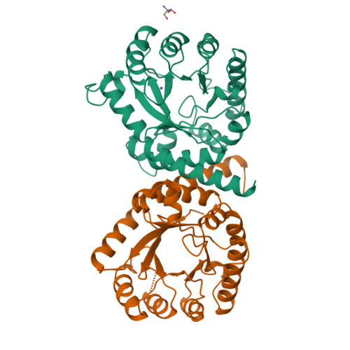
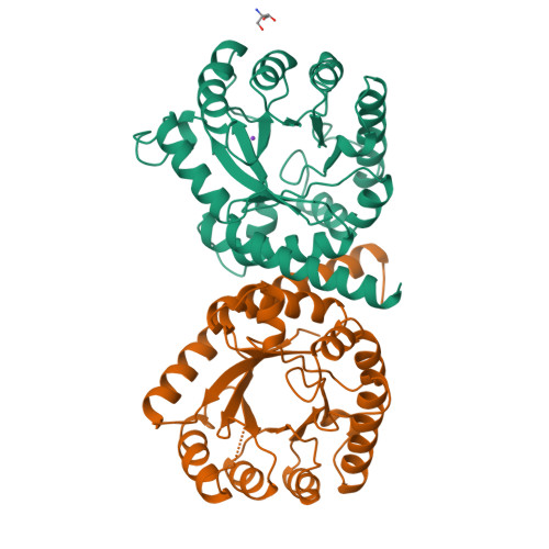
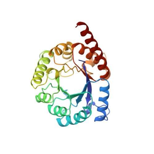
The gene encoding the dihydropteroate synthase of staphylococcus aureus has been cloned, sequenced and expressed in Escherichia coli. The protein has been purified for biochemical characterization and X-ray crystallographic studies. The enzyme is a dimer in solution, has a steady state kinetic mechanism that suggests random binding of the two substrates and half-site reactivity. The crystal structure of apo-enzyme and a binary complex with the substrate analogue hydroxymethylpterin pyrophosphate were determined at 2.2 A and 2.4 A resolution, respectively. The enzyme belongs to the group of "TIM-barrel" proteins and crystallizes as a non-crystallographic dimer. Only one molecule of the substrate analogue bound per dimer in the crystal. Sequencing of nine sulfonamide-resistant clinical isolates has shown that as many as 14 residues could be involved in resistance development. The residues are distributed over the surface of the protein, which defies a simple interpretation of their roles in resistance. Nevertheless, the three-dimensional structure of the substrate analogue binary complex could give important insight into the molecular mechanism of this enzyme.
Organizational Affiliation:
F. Hoffmann-La Roche Ltd, Pharma Preclinical Research Department, Basel, Switzerland.


