1AL7

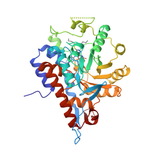
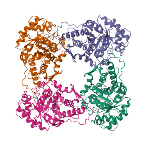
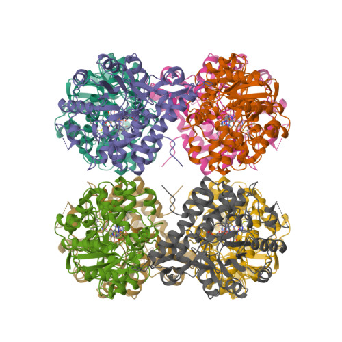
THREE-DIMENSIONAL STRUCTURES OF GLYCOLATE OXIDASE WITH BOUND ACTIVE-SITE INHIBITORS


Experimental Data Snapshot

  • Method: X-RAY DIFFRACTION
  • Resolution: 2.60 Å
  • R-Value Free: 0.242 
  • R-Value Work: 0.180 
  • R-Value Observed: 0.180 

wwPDB Validation   3D Report Full Report


This is version 1.3 of the entry. See complete history


Literature

Three-dimensional structures of glycolate oxidase with bound active-site inhibitors.

Stenberg, K.Lindqvist, Y.

(1997) Protein Sci 6: 1009-1015

  • DOI: https://doi.org/10.1002/pro.5560060506
  • Primary Citation of Related Structures:  
    1AL7, 1AL8

  • PubMed Abstract: 

    A key step in plant photorespiration, the oxidation of glycolate to glyoxylate, is carried out by the peroxisomal flavoprotein glycolate oxidase (EC 1.1.3.15). The three-dimensional structure of this alpha/beta barrel protein has been refined to 2 A resolution (Lindqvist Y. 1989. J Mol Biol 209:151-166). FMN dependent glycolate oxidase is a member of the family of alpha-hydroxy acid oxidases. Here we describe the crystallization and structure determination of two inhibitor complexes of the enzyme, TKP (3-Decyl-2,5-dioxo-4-hydroxy-3-pyrroline) and TACA (4-Carboxy-5-(1-pentyl)hexylsulfanyl-1,2,3-triazole). The structure of the TACA complex has been refined to 2.6 A resolution and the TKP complex, solved with molecular replacement, to 2.2 A resolution. The Rfree for the TACA and TKP complexes are 24.2 and 25.1%, respectively. The overall structures are very similar to the unliganded holoenzyme, but a closer examination of the active site reveals differences in the positioning of the flavin isoalloxazine ring and a displaced flexible loop in the TKP complex. The two inhibitors differ in binding mode and hydrophobic interactions, and these differences are reflected by the very different Ki values for the inhibitors, 16 nM for TACA and 4.8 microM for TKP. Implications of the structures of these enzyme-inhibitor complexes for the model for substrate binding and catalysis proposed from the holo-enzyme structure are discussed.


  • Organizational Affiliation

    Department of Medical Biochemistry and Biophysics, Karolinska, Institute, Stockholm, Sweden.


Macromolecules
Find similar proteins by:  (by identity cutoff)  |  3D Structure
Entity ID: 1
MoleculeChains Sequence LengthOrganismDetailsImage
GLYCOLATE OXIDASE359Spinacia oleraceaMutation(s): 0 
EC: 1.1.3.15
UniProt
Find proteins for P05414 (Spinacia oleracea)
Explore P05414 
Go to UniProtKB:  P05414
Entity Groups  
Sequence Clusters30% Identity50% Identity70% Identity90% Identity95% Identity100% Identity
UniProt GroupP05414
Sequence Annotations
Expand
  • Reference Sequence
Small Molecules
Ligands 2 Unique
IDChains Name / Formula / InChI Key2D Diagram3D Interactions
FMN
Query on FMN

Download Ideal Coordinates CCD File 
B [auth A]FLAVIN MONONUCLEOTIDE
C17 H21 N4 O9 P
FVTCRASFADXXNN-SCRDCRAPSA-N
HST
Query on HST

Download Ideal Coordinates CCD File 
C [auth A]4-CARBOXY-5-(1-PENTYL)HEXYLSULFANYL-1,2,3-TRIAZOLE
C14 H25 N3 O2 S
GROSWUGUHPUYIU-UHFFFAOYSA-N
Binding Affinity Annotations 
IDSourceBinding Affinity
HST PDBBind:  1AL7 Ki: 16 (nM) from 1 assay(s)
Experimental Data & Validation

Experimental Data

  • Method: X-RAY DIFFRACTION
  • Resolution: 2.60 Å
  • R-Value Free: 0.242 
  • R-Value Work: 0.180 
  • R-Value Observed: 0.180 
  • Space Group: I 4 2 2
Unit Cell:
Length ( Å )Angle ( ˚ )
a = 148.1α = 90
b = 148.1β = 90
c = 136.5γ = 90
Software Package:
Software NamePurpose
X-PLORmodel building
X-PLORrefinement
DENZOdata reduction
CCP4data scaling
X-PLORphasing

Structure Validation

View Full Validation Report


Entry History 

Deposition Data

Revision History  (Full details and data files)

  • Version 1.0: 1997-11-12
    Type: Initial release
  • Version 1.1: 2008-03-24
    Changes: Version format compliance
  • Version 1.2: 2011-07-13
    Changes: Derived calculations, Version format compliance
  • Version 1.3: 2024-02-07
    Changes: Data collection, Database references, Derived calculations, Other