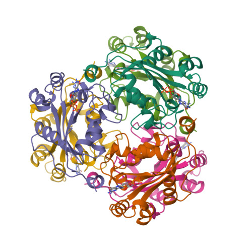
The three-dimensional structures of two isoforms of nucleoside diphosphate kinase from bovine retina.
Ladner, J.E., Abdulaev, N.G., Kakuev, D.L., Tordova, M., Ridge, K.D., Gilliland, G.L.(1999) Acta Crystallogr D Biol Crystallogr 55: 1127-1135
- PubMed: 10329774
- DOI: https://doi.org/10.1107/s0907444999002528
- Primary Citation of Related Structures:
1BHN - PubMed Abstract:
The crystal structures of two isoforms of nucleoside diphosphate kinase from bovine retina overexpressed in Escherischia coli have been determined to 2.4 A resolution. Both the isoforms, NBR-A and NBR-B, are hexameric and the fold of the monomer is in agreement with NDP-kinase structures from other biological sources. Although the polypeptide chains of the two isoforms differ by only two residues, they crystallize in different space groups. NBR-A crystallizes in space group P212121 with an entire hexamer in the asymmetric unit, while NBR-B crystallizes in space group P43212 with a trimer in the asymmetric unit. The highly conserved nucleotide-binding site observed in other nucleoside diphosphate kinase structures is also observed here. Both NBR-A and NBR-B were crystallized in the presence of cGMP. The nucleotide is bound with the base in the anti conformation. The NBR-A active site contained both cGMP and GDP each bound at half occupancy. Presumably, NBR-A had retained GDP (or GTP) from the purification process. The NBR-B active site contained only cGMP.
Organizational Affiliation:
Center for Advanced Research in Biotechnology, National Institute of Standards and Technology and the University of Maryland Biotechnology Institute, 9600 Gudelsky Drive, Rockville, MD 20850 USA.


