1ELQ | pdb_00001elq

CRYSTAL STRUCTURE OF THE CYSTINE C-S LYASE C-DES


Experimental Data Snapshot

  • Method: X-RAY DIFFRACTION
  • Resolution: 1.80 Å
  • R-Value Free: 
    0.249 (Depositor), 0.240 (DCC) 
  • R-Value Work: 
    0.198 (Depositor), 0.190 (DCC) 
  • R-Value Observed: 
    0.201 (Depositor) 

wwPDB Validation   3D Report Full Report


Ligand Structure Quality Assessment 

Created with Raphaël 2.3.0Worse 01 BetterLigand structure goodness of fit to experimental dataBest fitted PLPClick on this verticalbar to view details

This is version 1.3 of the entry. See complete history


Literature

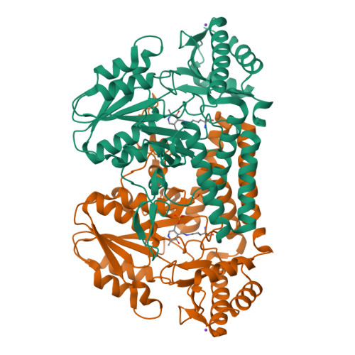
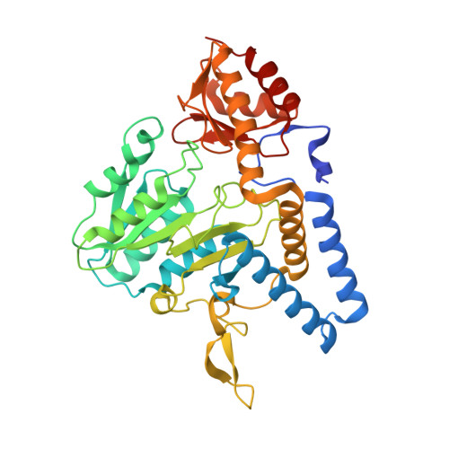
Crystal structure of the cystine C-S lyase from Synechocystis: stabilization of cysteine persulfide for FeS cluster biosynthesis.

Clausen, T.Kaiser, J.T.Steegborn, C.Huber, R.Kessler, D.

(2000) Proc Natl Acad Sci U S A 97: 3856-3861

  • DOI: https://doi.org/10.1073/pnas.97.8.3856
  • Primary Citation of Related Structures:  
    1ELQ, 1ELU

  • PubMed Abstract: 

    FeS clusters are versatile cofactors of a variety of proteins, but the mechanisms of their biosynthesis are still unknown. The cystine C-S lyase from Synechocystis has been identified as a participant in ferredoxin FeS cluster formation. Herein, we report on the crystal structure of the lyase and of a complex with the reaction products of cystine cleavage at 1.8- and 1.55-A resolution, respectively. The sulfur-containing product was unequivocally identified as cysteine persulfide. The reactive persulfide group is fixed by a hydrogen bond to His-114 in the center of a hydrophobic pocket and is thereby shielded from the solvent. Binding and stabilization of the cysteine persulfide represent an alternative to the generation of a protein-bound persulfide by NifS-like proteins and point to the general importance of persulfidic compounds for FeS cluster assembly.


  • Organizational Affiliation

    Max-Planck-Institut für Biochemie, Abteilung Strukturforschung, Am Klopferspitz 18a, D-82152 Martinsried, Germany. clausen@biochem.mpg.de


Macromolecules
Find similar proteins by:  (by identity cutoff)  |  3D Structure
Entity ID: 1
MoleculeChains Sequence LengthOrganismDetailsImage
L-CYSTEINE/L-CYSTINE C-S LYASE
A, B
390Synechocystis sp. PCC 6714Mutation(s): 8 
UniProt
Find proteins for Q9ZHG9 (Synechocystis sp. (strain PCC 6714))
Explore Q9ZHG9 
Go to UniProtKB:  Q9ZHG9
Entity Groups  
Sequence Clusters30% Identity50% Identity70% Identity90% Identity95% Identity100% Identity
UniProt GroupQ9ZHG9
Sequence Annotations
Expand
  • Reference Sequence
Experimental Data & Validation

Experimental Data

  • Method: X-RAY DIFFRACTION
  • Resolution: 1.80 Å
  • R-Value Free:  0.249 (Depositor), 0.240 (DCC) 
  • R-Value Work:  0.198 (Depositor), 0.190 (DCC) 
  • R-Value Observed: 0.201 (Depositor) 
Space Group: P 21 21 21
Unit Cell:
Length ( Å )Angle ( ˚ )
a = 62.4α = 90
b = 65.4β = 90
c = 170.1γ = 90
Software Package:
Software NamePurpose
SHARPphasing
CNSrefinement
DENZOdata reduction
CCP4data scaling

Structure Validation

View Full Validation Report


Ligand Structure Quality Assessment 

Created with Raphaël 2.3.0Worse 01 BetterLigand structure goodness of fit to experimental dataBest fitted PLPClick on this verticalbar to view details

Entry History 

Deposition Data

Revision History  (Full details and data files)

  • Version 1.0: 2000-04-19
    Type: Initial release
  • Version 1.1: 2008-04-27
    Changes: Version format compliance
  • Version 1.2: 2011-07-13
    Changes: Source and taxonomy, Version format compliance
  • Version 1.3: 2021-11-03
    Changes: Data collection, Database references, Derived calculations