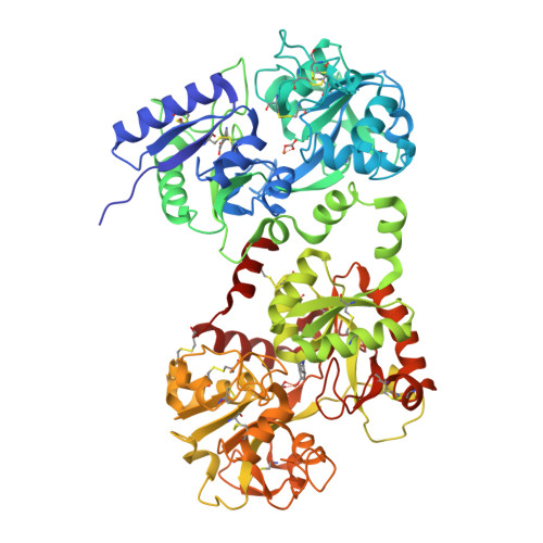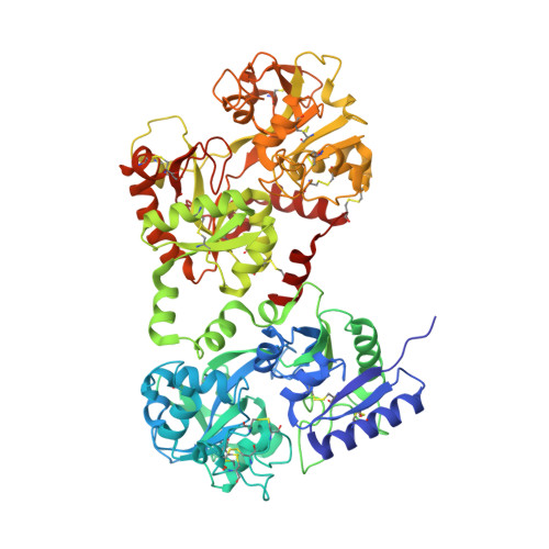
Lactoferrin-melanin interaction and its possible implications in melanin polymerization: crystal structure of the complex formed between mare lactoferrin and melanin monomers at 2.7-A resolution.
Sharma, A.K., Kumar, S., Sharma, V., Nagpal, A., Singh, N., Tamboli, I., Mani, I., Raman, G., Singh, T.P.(2001) Proteins 45: 229-236
- PubMed: 11599026
- DOI: https://doi.org/10.1002/prot.1143
- Primary Citation of Related Structures:
1F9B - PubMed Abstract:
The concentration of melanin determines the intensity of colors of the skin and hair of animals. Melanin pigments are tyrosine-based polymers formed in melanocytes within specialized organelles called melanosomes. In order to understand the mechanism of melanin polymerization, lactoferrin, a basic protein with a pI value of 9.0, has been used to produce melanin. Lactoferrin is a monomeric iron-binding protein with a molecular weight of 80 kDa. The crystals of lactoferrin were soaked in a solution containing dihydroxyphenylalanine (DOPA) and tyrosinase enzyme. These crystals were used for X-ray intensity data collection. The intensity data were collected to 2.7-A resolution to an overall completeness of 91% with an R(sym) of 0.071. The crystals belong to orthorhombic space group P2(1)2(1)2(1) with cell dimensions: a = 85.0 A, b = 99.8 A, c = 103.4 A. The structure was determined by molecular replacement method, using the model of diferric mare lactoferrin, and refined to an R-factor 0.215 (R(free) = 0.287) for all the data to 2.7-A resolution. The final model comprises 5,281 protein atoms from 689 amino acids, 2Fe(3+), 2CO(2-)(3) ions, 2 indole-5,6-quinone molecules (IQ), and 73 water molecules. Two IQ molecules, one in each lobe, bind to lactoferrin. In the C-lobe, the IQ binds in the iron-binding cleft, whereas in the N-lobe, it is located in the side pocket between two alpha-helices, filled with solvent molecules in the native iron-saturated mare lactoferrin. The IQ molecules interact with protein molecule mainly through glutamic acid in both lobes, without significant perturbation to the protein structure. The orientation of N- and C-lobes in the present structure is similar to that observed in the native iron-saturated protein. However, as a result of the binding of IQ molecules, the orientations of the domains N1, N2 and C1, C2 in the two cases differ slightly.
Organizational Affiliation:
Department of Biophysics, All India Institute of Medical Sciences, New Delhi, India.