1FDK | pdb_00001fdk

CARBOXYLIC ESTER HYDROLASE (PLA2-MJ33 INHIBITOR COMPLEX)


Experimental Data Snapshot

  • Method: X-RAY DIFFRACTION
  • Resolution: 1.91 Å
  • R-Value Free: 
    0.280 (Depositor) 
  • R-Value Work: 
    0.184 (Depositor), 0.200 (DCC) 
  • R-Value Observed: 
    0.184 (Depositor) 

Starting Model: experimental
View more details

wwPDB Validation   3D Report Full Report


Ligand Structure Quality Assessment 

Created with Raphaël 2.3.0Worse 01 BetterLigand structure goodness of fit to experimental dataBest fitted GLEClick on this verticalbar to view details

This is version 1.6 of the entry. See complete history


Literature

Crystal structure of the complex of bovine pancreatic phospholipase A2 with the inhibitor 1-hexadecyl-3-(trifluoroethyl)-sn-glycero-2-phosphomethanol,.

Sekar, K.Eswaramoorthy, S.Jain, M.K.Sundaralingam, M.

(1997) Biochemistry 36: 14186-14191

  • DOI: https://doi.org/10.1021/bi971370b
  • Primary Citation of Related Structures:  
    1FDK

  • PubMed Abstract: 

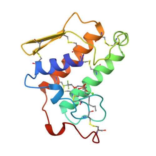
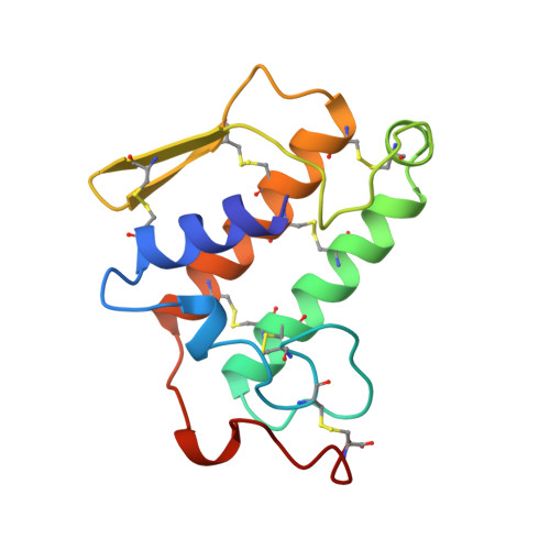
    The structure of recombinant bovine pancreatic phospholipase A2 (PLA2) complexed with the competitive inhibitor 1-hexadecyl-3-(trifluoroethyl)-sn-glycero -2-phosphomethanol (hereafter MJ33), a phospholipid analogue without the sn-3 phosphodiester group, has been determined. The crystals are trigonal, space group P3121, a = b = 46.36 A and c = 102.56 A, and isomorphous to the recombinant PLA2 with one molecule in the asymmetric unit. The structure was refined using 8082 reflections between 8.0 and 1.91 A resolution to a final R-value of 18.4% [Rfree = 28.0%]. The model includes 957 protein atoms, 86 water molecules, one calcium ion, and 26 non-hydrogen atoms of the inhibitor MJ33. The overall tertiary fold of the complex is very similar to that of the inhibitor-free recombinant PLA2 with a root mean square deviation of 0.32 A for all the backbone atoms. The electron density of the surface loop residues 62-66 is clear and ordered, unlike the other trigonal bovine PLA2 structures done to date. This structural change could be responsible for the interfacial allosteric activation, which thermodynamically relates the enhanced binding of the substrate mimic to the active site of the enzyme. MJ33 is tightly bound in the active-site cleft, dislodging the equatorial coordinated calcium water (W5), the putative catalytic water W6, and the neighboring water W7. The axial coordinated calcium water is missing; thus the hexacoordinated calcium is a monocapped pentagonal pyramid. Although MJ33 is a sn-2 tetrahedral mimic, its phosphate binds to PLA2 differently from the sn-2 phosphonate analogue of phospholipids, another tetrahedral mimic. The knowledge of the active-site geometry of MJ33 would be useful in the design of more useful therapeutic agents for PLA2.


  • Organizational Affiliation

    Biological Macromolecular Structure Center, Department of Chemistry, Ohio State University, Columbus, Ohio 43210, USA.


Macromolecules
Find similar proteins by:  (by identity cutoff)  |  3D Structure
Entity ID: 1
MoleculeChains Sequence LengthOrganismDetailsImage
PHOSPHOLIPASE A2123Bos taurusMutation(s): 0 
Gene Names: MATURE PLA2
EC: 3.1.1.4
UniProt
Find proteins for P00593 (Bos taurus)
Explore P00593 
Go to UniProtKB:  P00593
Entity Groups  
Sequence Clusters30% Identity50% Identity70% Identity90% Identity95% Identity100% Identity
UniProt GroupP00593
Sequence Annotations
Expand
  • Reference Sequence
Small Molecules
Experimental Data & Validation

Experimental Data

  • Method: X-RAY DIFFRACTION
  • Resolution: 1.91 Å
  • R-Value Free:  0.280 (Depositor) 
  • R-Value Work:  0.184 (Depositor), 0.200 (DCC) 
  • R-Value Observed: 0.184 (Depositor) 
Space Group: P 31 2 1
Unit Cell:
Length ( Å )Angle ( ˚ )
a = 46.36α = 90
b = 46.36β = 90
c = 102.56γ = 120
Software Package:
Software NamePurpose
R-AXISdata collection
R-AXISdata reduction
X-PLORmodel building
X-PLORrefinement
R-AXISdata scaling
X-PLORphasing

Structure Validation

View Full Validation Report


Ligand Structure Quality Assessment 

Created with Raphaël 2.3.0Worse 01 BetterLigand structure goodness of fit to experimental dataBest fitted GLEClick on this verticalbar to view details

Entry History 

Deposition Data

Revision History  (Full details and data files)

  • Version 1.0: 1998-10-14
    Type: Initial release
  • Version 1.1: 2008-03-24
    Changes: Version format compliance
  • Version 1.2: 2011-07-13
    Changes: Version format compliance
  • Version 1.3: 2018-04-04
    Changes: Data collection, Other
  • Version 1.4: 2018-04-11
    Changes: Data collection
  • Version 1.5: 2024-04-03
    Changes: Data collection, Database references, Derived calculations, Refinement description
  • Version 1.6: 2024-10-30
    Changes: Structure summary