1G9V | pdb_00001g9v

HIGH RESOLUTION CRYSTAL STRUCTURE OF DEOXY HEMOGLOBIN COMPLEXED WITH A POTENT ALLOSTERIC EFFECTOR


Experimental Data Snapshot

  • Method: X-RAY DIFFRACTION
  • Resolution: 1.85 Å
  • R-Value Free: 
    0.208 (Depositor) 
  • R-Value Work: 
    0.177 (Depositor), 0.170 (DCC) 
  • R-Value Observed: 
    0.179 (Depositor) 

Starting Model: experimental
View more details

wwPDB Validation   3D Report Full Report


Ligand Structure Quality Assessment 

Created with Raphaël 2.3.0Worse 01 BetterLigand structure goodness of fit to experimental dataBest fitted HEMClick on this verticalbar to view detailsBest fitted RQ3Click on this verticalbar to view details

This is version 1.4 of the entry. See complete history


Literature

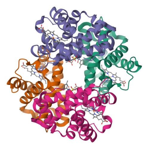
High-resolution crystal structure of deoxy hemoglobin complexed with a potent allosteric effector.

Safo, M.K.Moure, C.M.Burnett, J.C.Joshi, G.S.Abraham, D.J.

(2001) Protein Sci 10: 951-957

  • DOI: https://doi.org/10.1110/ps.50601
  • Primary Citation of Related Structures:  
    1G9V

  • PubMed Abstract: 

    The crystal structure of human deoxy hemoglobin (Hb) complexed with a potent allosteric effector (2-[4-[[(3,5-dimethylanilino)carbonyl]methyl]phenoxy]-2-methylpropionic acid) = RSR-13) is reported at 1.85 A resolution. Analysis of the hemoglobin:effector complex indicates that two of these molecules bind to the central water cavity of deoxy Hb in a symmetrical fashion, and that each constrains the protein by engaging in hydrogen bonding and hydrophobic interactions with three of its four subunits. Interestingly, we also find that water-mediated interactions between the bound effectors and the protein make significant contributions to the overall binding. Physiologically, the interaction of RSR-13 with Hb results in increased oxygen delivery to peripheral tissues. Thus, this compound has potential therapeutic application in the treatment of hypoxia, ischemia, and trauma-related blood loss. Currently, RSR-13 is in phase III clinical trials as a radiosensitizing agent in the treatment of brain tumors. A detailed structural analysis of this compound complexed with deoxy Hb has important implications for the rational design of future analogs.


  • Organizational Affiliation

    Department of Medicinal Chemistry, School of Pharmacy and Institute for Structural Biology and Drug Discovery, Virginia Commonwealth University, Richmond, Virginia 23298-0540, USA.


Macromolecules
Find similar proteins by:  (by identity cutoff)  |  3D Structure
Entity ID: 1
MoleculeChains Sequence LengthOrganismDetailsImage
HEMOGLOBIN ALPHA CHAIN
A, C
141Homo sapiensMutation(s): 0 
UniProt & NIH Common Fund Data Resources
Find proteins for P69905 (Homo sapiens)
Explore P69905 
Go to UniProtKB:  P69905
PHAROS:  P69905
Entity Groups  
Sequence Clusters30% Identity50% Identity70% Identity90% Identity95% Identity100% Identity
UniProt GroupP69905
Sequence Annotations
Expand
  • Reference Sequence
Find similar proteins by:  (by identity cutoff)  |  3D Structure
Entity ID: 2
MoleculeChains Sequence LengthOrganismDetailsImage
HEMOGLOBIN BETA CHAIN
B, D
146Homo sapiensMutation(s): 0 
UniProt & NIH Common Fund Data Resources
Find proteins for P68871 (Homo sapiens)
Explore P68871 
Go to UniProtKB:  P68871
PHAROS:  P68871
GTEx:  ENSG00000244734 
Entity Groups  
Sequence Clusters30% Identity50% Identity70% Identity90% Identity95% Identity100% Identity
UniProt GroupP68871
Sequence Annotations
Expand
  • Reference Sequence
Small Molecules
Ligands 3 Unique
IDChains Name / Formula / InChI Key2D Diagram3D Interactions
HEM
Query on HEM

Download Ideal Coordinates CCD File 
E [auth A],
H [auth B],
I [auth C],
L [auth D]
PROTOPORPHYRIN IX CONTAINING FE
C34 H32 Fe N4 O4
KABFMIBPWCXCRK-RGGAHWMASA-L
RQ3
Query on RQ3

Download Ideal Coordinates CCD File 
F [auth A],
J [auth C]
2-{4-[(3,5-DIMETHYLANILINO)-CARBONYL-METHYL]-PHENOXY}-2-METHYLPROPIONIC ACID
C20 H23 N O4
BNFRJXLZYUTIII-UHFFFAOYSA-N
SO4
Query on SO4

Download Ideal Coordinates CCD File 
G [auth B],
K [auth D]
SULFATE ION
O4 S
QAOWNCQODCNURD-UHFFFAOYSA-L
Experimental Data & Validation

Experimental Data

  • Method: X-RAY DIFFRACTION
  • Resolution: 1.85 Å
  • R-Value Free:  0.208 (Depositor) 
  • R-Value Work:  0.177 (Depositor), 0.170 (DCC) 
  • R-Value Observed: 0.179 (Depositor) 
Space Group: P 1 21 1
Unit Cell:
Length ( Å )Angle ( ˚ )
a = 63.2α = 90
b = 83.56β = 99.16
c = 53.86γ = 90
Software Package:
Software NamePurpose
X-PLORmodel building
X-PLORrefinement
CCP4data scaling
ROTAVATAdata scaling
TRUNCATEdata scaling
X-PLORphasing

Structure Validation

View Full Validation Report


Ligand Structure Quality Assessment 

Created with Raphaël 2.3.0Worse 01 BetterLigand structure goodness of fit to experimental dataBest fitted HEMClick on this verticalbar to view detailsBest fitted RQ3Click on this verticalbar to view details

Entry History 

Deposition Data

Revision History  (Full details and data files)

  • Version 1.0: 2000-12-06
    Type: Initial release
  • Version 1.1: 2008-04-27
    Changes: Version format compliance
  • Version 1.2: 2011-07-13
    Changes: Version format compliance
  • Version 1.3: 2017-10-04
    Changes: Refinement description
  • Version 1.4: 2023-08-09
    Changes: Data collection, Database references, Derived calculations, Refinement description