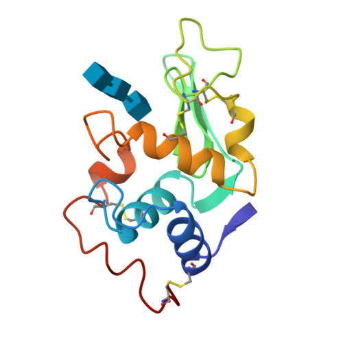
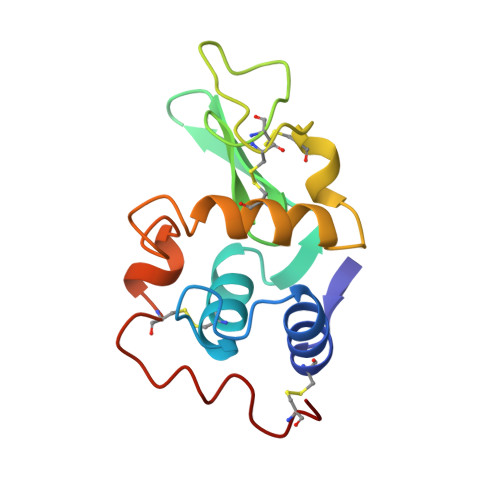
1HEW

REFINEMENT OF AN ENZYME COMPLEX WITH INHIBITOR BOUND AT PARTIAL OCCUPANCY. HEN EGG-WHITE LYSOZYME AND TRI-N-ACETYLCHITOTRIOSE AT 1.75 ANGSTROMS RESOLUTION


Experimental Data Snapshot

  • Method: X-RAY DIFFRACTION
  • Resolution: 1.75 Å
  • R-Value Observed: 0.229 

wwPDB Validation   3D Report Full Report


This is version 2.1 of the entry. See complete history


Literature

Refinement of an enzyme complex with inhibitor bound at partial occupancy. Hen egg-white lysozyme and tri-N-acetylchitotriose at 1.75 A resolution.

Cheetham, J.C.Artymiuk, P.J.Phillips, D.C.

(1992) J Mol Biol 224: 613-628

  • DOI: https://doi.org/10.1016/0022-2836(92)90548-x
  • Primary Citation of Related Structures:  
    1HEW

  • PubMed Abstract: 

    The structure of the tri-N-acetylchitotriose inhibitor complex of hen egg-white lysozyme has been refined at 1.75 A resolution, using data collected from a complex crystal with ligand bound at less than full occupancy. To determine the exact value of the inhibitor occupancy, a model comprising unliganded and sugar-bound protein molecules was generated and refined against the 1.75 A data, using a modified version of the Hendrickson & Konnert least-squares procedure. The crystallographic R-factor for the model was found to fall to a minimum at 55% bound sugar. Conventional refinement assuming unit occupancy was found to yield incorrect thermal and positional parameters. Application of the same refinement procedures to an earlier 2.0 A data set, collected independently on different complex crystals by Blake et al. gave less consistent results than the 1.75 A refinement. From an analysis of the high resolution structure a detailed picture of the protein-carbohydrate interactions in the non-productive complex has emerged, together with the conformation and mobility changes that accompany ligand binding. The specificity of interaction between the protein and inhibitor, bound in subsites A to C of the active site, is seen to be generated primarily by an extensive network of hydrogen bonds, both to the protein itself and to bound solvent molecules. The latter also play an important role in maintaining the structural integrity of the active site cleft in the apo-protein.


  • Organizational Affiliation

    Laboratory of Molecular Biophysics, University of Oxford, England.


Macromolecules
Find similar proteins by:  (by identity cutoff)  |  3D Structure
Entity ID: 1
MoleculeChains Sequence LengthOrganismDetailsImage
HEN EGG WHITE LYSOZYME129Gallus gallusMutation(s): 0 
EC: 3.2.1.17
UniProt
Find proteins for P00698 (Gallus gallus)
Explore P00698 
Go to UniProtKB:  P00698
Entity Groups  
Sequence Clusters30% Identity50% Identity70% Identity90% Identity95% Identity100% Identity
UniProt GroupP00698
Sequence Annotations
Expand
  • Reference Sequence
Oligosaccharides

Help

Entity ID: 2
MoleculeChains Length2D Diagram Glycosylation3D Interactions
2-acetamido-2-deoxy-beta-D-glucopyranose-(1-4)-2-acetamido-2-deoxy-beta-D-glucopyranose-(1-4)-2-acetamido-2-deoxy-beta-D-glucopyranose
B
3N/A
Glycosylation Resources
GlyTouCan:  G47362BJ
GlyCosmos:  G47362BJ
GlyGen:  G47362BJ
Biologically Interesting Molecules (External Reference) 1 Unique
Experimental Data & Validation

Experimental Data

  • Method: X-RAY DIFFRACTION
  • Resolution: 1.75 Å
  • R-Value Observed: 0.229 
  • Space Group: P 43 21 2
Unit Cell:
Length ( Å )Angle ( ˚ )
a = 78.86α = 90
b = 78.86β = 90
c = 38.25γ = 90
Software Package:
Software NamePurpose
PROLSQrefinement

Structure Validation

View Full Validation Report


Entry History 

Deposition Data

Revision History  (Full details and data files)

  • Version 1.0: 1994-01-31
    Type: Initial release
  • Version 1.1: 2008-03-24
    Changes: Version format compliance
  • Version 1.2: 2011-07-13
    Changes: Non-polymer description, Version format compliance
  • Version 1.3: 2017-11-29
    Changes: Derived calculations, Other
  • Version 2.0: 2020-07-29
    Type: Remediation
    Reason: Carbohydrate remediation
    Changes: Atomic model, Data collection, Derived calculations, Structure summary
  • Version 2.1: 2024-10-30
    Changes: Data collection, Database references, Structure summary