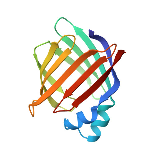
Escherichia coli-derived rat intestinal fatty acid binding protein with bound myristate at 1.5 A resolution and I-FABPArg106-->Gln with bound oleate at 1.74 A resolution.
Eads, J., Sacchettini, J.C., Kromminga, A., Gordon, J.I.(1993) J Biol Chem 268: 26375-26385
- PubMed: 8253762
- Primary Citation of Related Structures:
1ICM, 1ICN - PubMed Abstract:
Rat intestinal fatty acid binding protein (I-FABP) is a 131-residue protein composed of two short alpha-helices (alpha I and alpha II) and 10 anti-parallel beta-strands organized into two nearly orthogonal beta-sheets. The structure of crystalline I-FABP with bound tetradecanoate (myristate) has been refined to a resolution of 1.5 A and compared to the 1.2 A structure of apo-I-FABP, the 1.9 A structure of I-FABP:hexadecanoate (palmitate) and the 1.75 A structure of I-FABP:9Z-octadecanoate (oleate) to determine how this model fatty acid receptor accommodates changes in the length of its fatty acid ligand. Myristate is located in the interior of the protein. A highly ordered, electrostatic network containing 7 hydrogen (H)-bonds links the OE1 and OE2 atoms of myristate's carboxylate group, the indole nitrogen of Trp82, NH1, and NH2 of Arg106, NE2, and OE1 of Gln115, and 2 interior ordered waters. The hydrocarbon chain of the bound fatty acid is slightly bent. Its convex face lies in a crevice, forming van der Waals contacts with the side chains of several hydrophobic and aromatic residues. Its concave face is exposed to an array of 8 interior ordered waters whose positions are stabilized by H-bond interactions with other waters, H-bond interactions with the side chains of polar/ionizable residues, and van der Waals contacts with the surface of the fatty acid. Addition of 2 or 4 methylenes to myristate produces remarkably little change in the positions of I-FABP's main chain and side chain atoms and interior ordered waters. The principal alterations are in the conformation of a surface opening (portal) connecting external and internal solvent and in the position of the benzene side chain of Phe55. Changes in the conformation of the portal reflect movement of two of its components: the backbone of alpha II and a type I turn (Ala73, Asp74) connecting two beta-strands. The positions of the main chain atoms of Ala73 and Asp74 appear to be determined by their ability to form van der Waals contacts with the omega-terminus of the fatty acid. The side chain of Phe55 appears to function as an adjustable aromatic lid, located over the portal, whose position is dependent on an ability to form van der Waals contacts with a fatty acid's omega-terminus.(ABSTRACT TRUNCATED AT 400 WORDS)
Organizational Affiliation:
Department of Biochemistry, Albert Einstein College of Medicine, Bronx, New York 10461.


