1IO3

CRYSTAL STRUCTURE OF FERRICYTOCHROME C2 FROM RHODOPSEUDOMONAS VIRIDIS


Experimental Data Snapshot

  • Method: X-RAY DIFFRACTION
  • Resolution: 1.90 Å
  • R-Value Free: 0.243 
  • R-Value Work: 0.208 

Starting Model: experimental
View more details

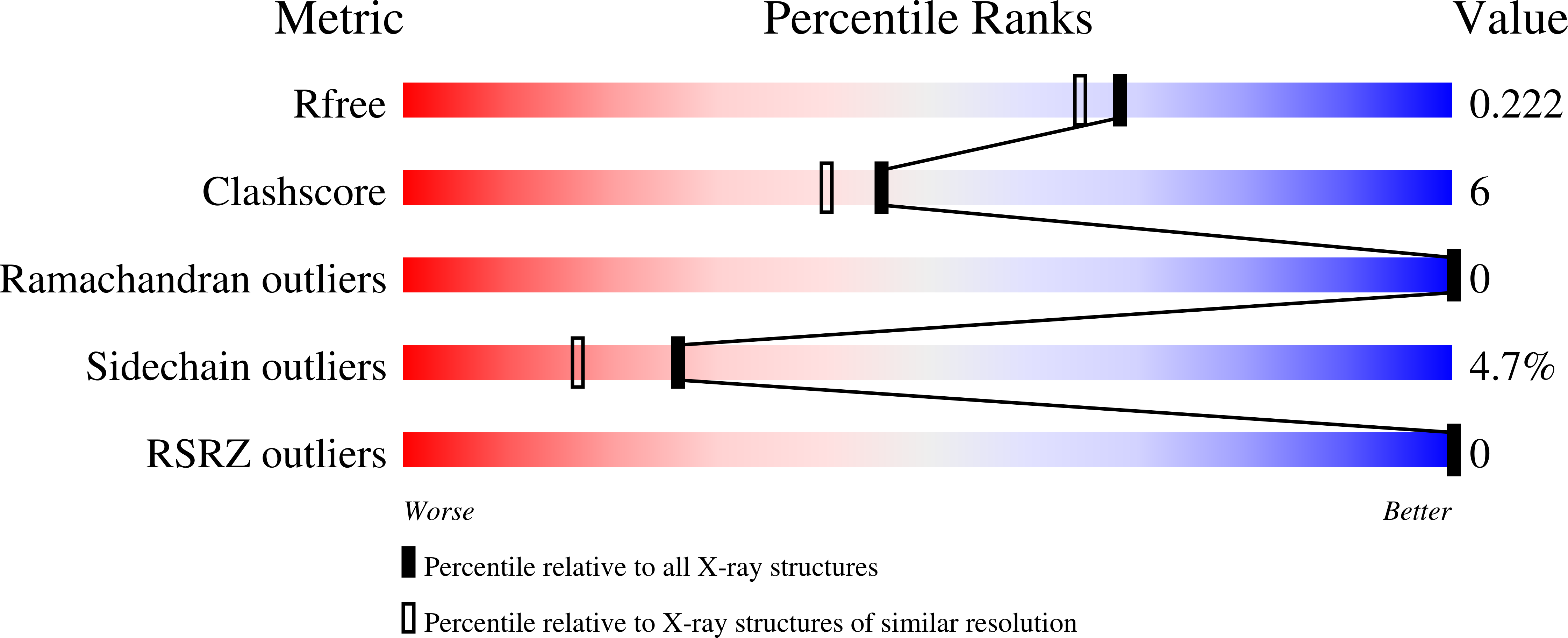
wwPDB Validation   3D Report Full Report


Ligand Structure Quality Assessment 


This is version 2.2 of the entry. See complete history


Literature

Crystal structure of the oxidized cytochrome c(2) from Blastochloris viridis.

Sogabe, S.Miki, K.

(2001) FEBS Lett 491: 174-179

  • DOI: https://doi.org/10.1016/s0014-5793(01)02179-2
  • Primary Citation of Related Structures:  
    1IO3

  • PubMed Abstract: 

    The crystal structure of the oxidized cytochrome c(2) from Blastochloris (formerly Rhodopseudomonas) viridis was determined at 1.9 A resolution. Structural comparison with the reduced form revealed significant structural changes according to the oxidation state of the heme iron. Slight perturbation of the polypeptide chain backbone was observed, and the secondary structure and the hydrogen patterns between main-chain atoms were retained. The oxidation state-dependent conformational shifts were localized in the vicinity of the methionine ligand side and the propionate group of the heme. The conserved segment of the polypeptide chain in cytochrome c and cytochrome c(2) exhibited some degree of mobility, interacting with the heme iron atom by the hydrogen bond network. These results indicate that the movement of the internal water molecule conserved in various c-type cytochromes drives the adjustments of side-chain atoms of nearby residue, and the segmental temperature factor changes along the polypeptide chain.


  • Organizational Affiliation

    Research Laboratory of Resources Utilization, Tokyo Institute of Technology, Yokohama, Japan.


Macromolecules
Find similar proteins by:  (by identity cutoff)  |  3D Structure
Entity ID: 1
MoleculeChains Sequence LengthOrganismDetailsImage
CYTOCHROME C2107Blastochloris viridisMutation(s): 0 
UniProt
Find proteins for P00083 (Blastochloris viridis)
Explore P00083 
Go to UniProtKB:  P00083
Entity Groups  
Sequence Clusters30% Identity50% Identity70% Identity90% Identity95% Identity100% Identity
UniProt GroupP00083
Sequence Annotations
Expand
  • Reference Sequence
Small Molecules
Ligands 1 Unique
IDChains Name / Formula / InChI Key2D Diagram3D Interactions
HEC
Query on HEC

Download Ideal Coordinates CCD File 
B [auth A]HEME C
C34 H34 Fe N4 O4
HXQIYSLZKNYNMH-LJNAALQVSA-N
Experimental Data & Validation

Experimental Data

  • Method: X-RAY DIFFRACTION
  • Resolution: 1.90 Å
  • R-Value Free: 0.243 
  • R-Value Work: 0.208 
  • Space Group: P 32 2 1
Unit Cell:
Length ( Å )Angle ( ˚ )
a = 75.97α = 90
b = 75.97β = 90
c = 40.37γ = 120
Software Package:
Software NamePurpose
AMoREphasing
X-PLORrefinement
DENZOdata reduction
SCALEPACKdata scaling

Structure Validation

View Full Validation Report


Ligand Structure Quality Assessment 


Entry History 

Deposition Data

Revision History  (Full details and data files)

  • Version 1.0: 2001-04-18
    Type: Initial release
  • Version 1.1: 2008-04-27
    Changes: Version format compliance
  • Version 1.2: 2011-07-13
    Changes: Version format compliance
  • Version 2.0: 2019-10-02
    Changes: Atomic model, Data collection, Derived calculations, Non-polymer description, Structure summary
  • Version 2.1: 2023-10-25
    Changes: Data collection, Database references, Refinement description
  • Version 2.2: 2024-10-23
    Changes: Structure summary