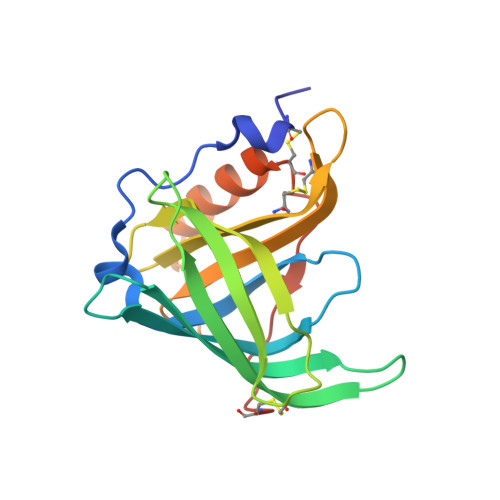
Role of conserved residues in structure and stability: Tryptophans of human serum retinol-binding protein, a model for the lipocalin superfamily
Greene, L.H., Chrysina, E.D., Irons, L.I., Papageorgiou, A.C., Acharya, K.R., Brew, K.(2001) Protein Sci 10: 2301-2316
- PubMed: 11604536
- DOI: https://doi.org/10.1110/ps.22901
- Primary Citation of Related Structures:
1JYD, 1JYJ - PubMed Abstract:
Serum retinol binding protein (RBP) is a member of the lipocalin family, proteins with up-and-down beta-barrel folds, low levels of sequence identity, and diverse functions. Although tryptophan 24 of RBP is highly conserved among lipocalins, it does not play a direct role in activity. To determine if Trp24 and other conserved residues have roles in stability and/or folding, we investigated the effects of conservative substitutions for the four tryptophans and some adjacent residues on the structure, stability, and spectroscopic properties of apo-RBP. Crystal structures of recombinant human apo-RBP and of a mutant with substitutions for tryptophans 67 and 91 at 1.7 A and 2.0 A resolution, respectively, as well as stability measurements, indicate that these relatively exposed tryptophans have little influence on structure or stability. Although Trp105 is largely buried in the wall of the beta-barrel, it can be replaced with minor effects on stability to thermal and chemical unfolding. In contrast, substitutions of three different amino acids for Trp24 or replacement of Arg139, a conserved residue that interacts with Trp24, lead to similar large losses in stability and lower yields of native protein generated by in vitro folding. The results and the coordinated nature of natural substitutions at these sites support the idea that conserved residues in functionally divergent homologs have roles in stabilizing the native relative to misfolded structures. They also establish conditions for studies of the kinetics of folding and unfolding by identifying spectroscopic signals for monitoring the formation of different substructures.
Organizational Affiliation:
Department of Biochemistry and Molecular Biology, University of Miami School of Medicine, Miami, Florida 33101, USA.


