1K3U | pdb_00001k3u

CRYSTAL STRUCTURE OF WILD-TYPE TRYPTOPHAN SYNTHASE COMPLEXED WITH N-[1H-INDOL-3-YL-ACETYL]ASPARTIC ACID


Experimental Data Snapshot

  • Method: X-RAY DIFFRACTION
  • Resolution: 1.70 Å
  • R-Value Free: 
    0.188 (Depositor), 0.190 (DCC) 
  • R-Value Work: 
    0.153 (Depositor), 0.160 (DCC) 
  • R-Value Observed: 
    0.155 (Depositor) 

Starting Model: experimental
View more details

wwPDB Validation   3D Report Full Report


Ligand Structure Quality Assessment 

Created with Raphaël 2.3.0Worse 01 BetterLigand structure goodness of fit to experimental dataBest fitted IADClick on this verticalbar to view detailsBest fitted PLPClick on this verticalbar to view details

This is version 1.4 of the entry. See complete history


Literature

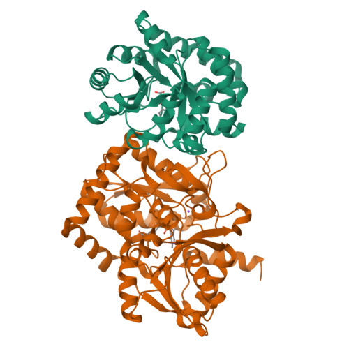
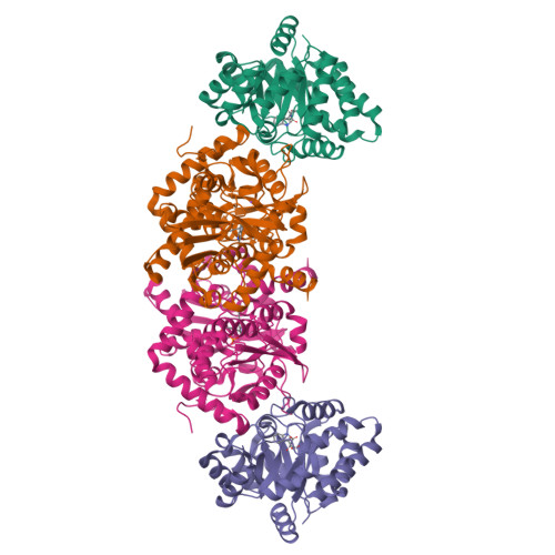
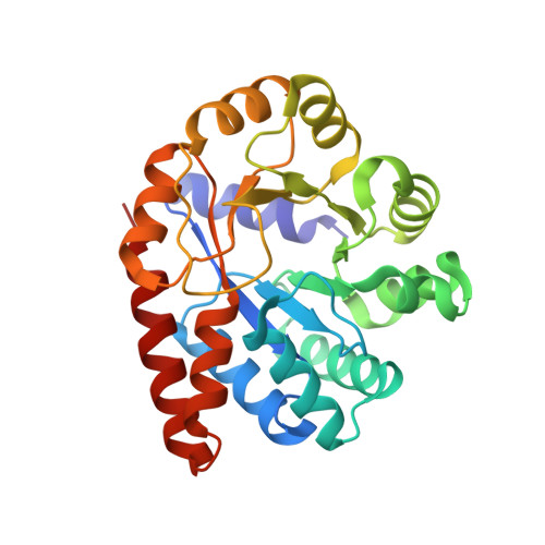
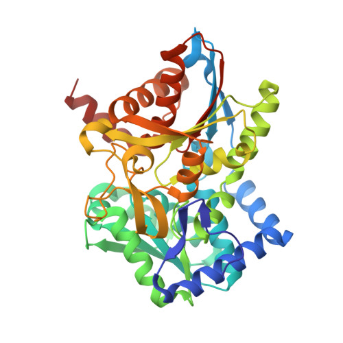
Crystal structures of a new class of allosteric effectors complexed to tryptophan synthase.

Weyand, M.Schlichting, I.Marabotti, A.Mozzarelli, A.

(2002) J Biological Chem 277: 10647-10652

  • DOI: https://doi.org/10.1074/jbc.M111285200
  • Primary Citation of Related Structures:  
    1K3U, 1K7E, 1K7F

  • PubMed Abstract: 

    Tryptophan synthase is a bifunctional alpha(2)beta(2) complex catalyzing the last two steps of l-tryptophan biosynthesis. The natural substrates of the alpha-subunit indole- 3-glycerolphosphate and glyceraldehyde-3-phosphate, and the substrate analogs indole-3-propanolphosphate and dl-alpha-glycerol-3-phosphate are allosteric effectors of the beta-subunit activity. It has been shown recently, that the indole-3-acetyl amino acids indole-3-acetylglycine and indole-3-acetyl-l-aspartic acid are both alpha-subunit inhibitors and beta-subunit allosteric effectors, whereas indole-3-acetyl-l-valine is only an alpha-subunit inhibitor (Marabotti, A., Cozzini, P., and Mozzarelli, A. (2000) Biochim. Biophys. Acta 1476, 287-299). The crystal structures of tryptophan synthase complexed with indole-3-acetylglycine and indole-3-acetyl-l-aspartic acid show that both ligands bind to the active site such that the carboxylate moiety is positioned similarly as the phosphate group of the natural substrates. As a consequence, the residues of the alpha-active site that interact with the ligands are the same as observed in the indole 3-glycerolphosphate-enzyme complex. Ligand binding leads to closure of loop alphaL6 of the alpha-subunit, a key structural element of intersubunit communication. This is in keeping with the allosteric role played by these compounds. The structure of the enzyme complex with indole-3-acetyl-l-valine is quite different. Due to the hydrophobic lateral chain, this molecule adopts a new orientation in the alpha-active site. In this case, closure of loop alphaL6 is no longer observed, in agreement with its functioning only as an inhibitor of the alpha-subunit reaction.


  • Organizational Affiliation

    Max-Planck-Institut für Molekulare Physiologie, Abteilung für Physikalische Biochemie, D-44227 Dortmund, Germany.


Macromolecules
Find similar proteins by:  (by identity cutoff)  |  3D Structure
Entity ID: 1
MoleculeChains Sequence LengthOrganismDetailsImage
Tryptophan Synthase Alpha Chain268Salmonella enterica subsp. enterica serovar TyphimuriumMutation(s): 0 
Gene Names: TRPA/TRPB
EC: 4.2.1.20
UniProt
Find proteins for P00929 (Salmonella typhimurium (strain LT2 / SGSC1412 / ATCC 700720))
Explore P00929 
Go to UniProtKB:  P00929
Entity Groups  
Sequence Clusters30% Identity50% Identity70% Identity90% Identity95% Identity100% Identity
UniProt GroupP00929
Sequence Annotations
Expand
  • Reference Sequence
Find similar proteins by:  (by identity cutoff)  |  3D Structure
Entity ID: 2
MoleculeChains Sequence LengthOrganismDetailsImage
Tryptophan Synthase Beta Chain396Salmonella enterica subsp. enterica serovar TyphimuriumMutation(s): 0 
Gene Names: TRPA/TRPB
EC: 4.2.1.20
UniProt
Find proteins for P0A2K1 (Salmonella typhimurium (strain LT2 / SGSC1412 / ATCC 700720))
Explore P0A2K1 
Go to UniProtKB:  P0A2K1
Entity Groups  
Sequence Clusters30% Identity50% Identity70% Identity90% Identity95% Identity100% Identity
UniProt GroupP0A2K1
Sequence Annotations
Expand
  • Reference Sequence
Experimental Data & Validation

Experimental Data

  • Method: X-RAY DIFFRACTION
  • Resolution: 1.70 Å
  • R-Value Free:  0.188 (Depositor), 0.190 (DCC) 
  • R-Value Work:  0.153 (Depositor), 0.160 (DCC) 
  • R-Value Observed: 0.155 (Depositor) 
Space Group: C 1 2 1
Unit Cell:
Length ( Å )Angle ( ˚ )
a = 182.409α = 90
b = 59.718β = 94.52
c = 67.177γ = 90
Software Package:
Software NamePurpose
REFMACrefinement
CNSrefinement
XDSdata reduction
XSCALEdata scaling
CNSphasing

Structure Validation

View Full Validation Report


Ligand Structure Quality Assessment 

Created with Raphaël 2.3.0Worse 01 BetterLigand structure goodness of fit to experimental dataBest fitted IADClick on this verticalbar to view detailsBest fitted PLPClick on this verticalbar to view details

Entry History 

Deposition Data

Revision History  (Full details and data files)

  • Version 1.0: 2002-07-03
    Type: Initial release
  • Version 1.1: 2008-04-27
    Changes: Version format compliance
  • Version 1.2: 2011-07-13
    Changes: Non-polymer description, Version format compliance
  • Version 1.3: 2019-07-24
    Changes: Data collection, Derived calculations, Refinement description
  • Version 1.4: 2023-08-16
    Changes: Data collection, Database references, Derived calculations, Refinement description