RCSB PDB - 1KNK: Crystal Structure of 2-C-methyl-D-erythritol 2,4-cyclodiphosphate Synthase (ispF) from E. coli involved in Mevalonate-Independent Isoprenoid Biosynthesis

 1KNK

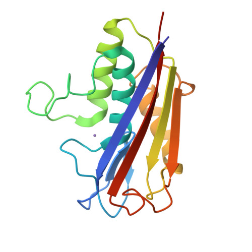
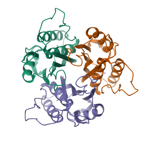
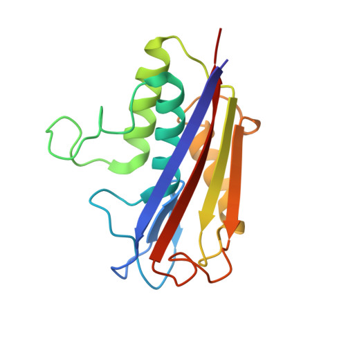
Crystal Structure of 2-C-methyl-D-erythritol 2,4-cyclodiphosphate Synthase (ispF) from E. coli involved in Mevalonate-Independent Isoprenoid Biosynthesis


Experimental Data Snapshot

  • Method: X-RAY DIFFRACTION
  • Resolution: 2.80 Å
  • R-Value Free: 0.249 
  • R-Value Work: 0.232 

Starting Model: experimental
View more details

wwPDB Validation   3D Report Full Report


This is version 1.4 of the entry. See complete history


Literature

Structure and mechanism of 2-C-methyl-D-erythritol 2,4-cyclodiphosphate synthase. An enzyme in the mevalonate-independent isoprenoid biosynthetic pathway.

Richard, S.B.Ferrer, J.L.Bowman, M.E.Lillo, A.M.Tetzlaff, C.N.Cane, D.E.Noel, J.P.

(2002) J Biol Chem 277: 8667-8672

  • DOI: https://doi.org/10.1074/jbc.C100739200
  • Primary Citation of Related Structures:  
    1KNJ, 1KNK

  • PubMed Abstract: 

    The enzyme 2-C-methyl-D-erythritol 2,4-cyclodiphosphate (MECDP) synthase catalyzes the conversion of 4-diphosphocytidyl-2-C-methyl-D-erythritol 2-phosphate (CDP-ME2P) to MECDP, a highly unusual cyclodiphosphate-containing intermediate on the mevalonate-independent pathway to isopentenyl diphosphate and dimethylallyl diphosphate. We now report two x-ray crystal structures of MECDP synthase refined to 2.8-A resolution. The first structure contains a bound Mn(2+) cation, and the second structure contains CMP, MECDP, and Mn(2+). The protein adopts a homotrimeric quaternary structure built around a central hydrophobic cavity and three externally facing active sites. Each of these active sites is located between two adjacent monomers. A tetrahedrally arranged transition metal binding site, potentially occupied by Mn(2+), sits at the base of the active site cleft. A phosphate oxygen of MECDP and the side chains of Asp(8), His(10), and His(42) occupy the metal ion coordination sphere. These structures reveal for the first time the structural determinants underlying substrate, product, and Mn(2+) recognition and the likely catalytic mechanism accompanying the biosynthesis of the cyclodiphosphate-containing isoprenoid precursor, MECDP.


  • Organizational Affiliation

    Structural Biology Laboratory, The Salk Institute for Biological Studies, La Jolla, California 92037, USA.


Macromolecules
Find similar proteins by:  (by identity cutoff)  |  3D Structure
Entity ID: 1
MoleculeChains Sequence LengthOrganismDetailsImage
2C-methyl-D-erythritol 2,4-cyclodiphosphate synthase159Escherichia coliMutation(s): 0 
Gene Names: ispF
EC: 4.6.1.12
UniProt
Find proteins for P62617 (Escherichia coli (strain K12))
Explore P62617 
Go to UniProtKB:  P62617
Entity Groups  
Sequence Clusters30% Identity50% Identity70% Identity90% Identity95% Identity100% Identity
UniProt GroupP62617
Sequence Annotations
Expand
  • Reference Sequence
Small Molecules
Ligands 1 Unique
IDChains Name / Formula / InChI Key2D Diagram3D Interactions
MN
Query on MN

Download Ideal Coordinates CCD File 
B [auth A]MANGANESE (II) ION
Mn
WAEMQWOKJMHJLA-UHFFFAOYSA-N
Experimental Data & Validation

Experimental Data

  • Method: X-RAY DIFFRACTION
  • Resolution: 2.80 Å
  • R-Value Free: 0.249 
  • R-Value Work: 0.232 
  • Space Group: I 21 3
Unit Cell:
Length ( Å )Angle ( ˚ )
a = 145.63α = 90
b = 145.63β = 90
c = 145.63γ = 90
Software Package:
Software NamePurpose
CNSrefinement
DENZOdata reduction
SCALEPACKdata scaling
CNSphasing

Structure Validation

View Full Validation Report


Entry History 

Deposition Data

Revision History  (Full details and data files)

  • Version 1.0: 2002-06-18
    Type: Initial release
  • Version 1.1: 2008-04-27
    Changes: Version format compliance
  • Version 1.2: 2011-07-13
    Changes: Derived calculations, Version format compliance
  • Version 1.3: 2011-11-16
    Changes: Atomic model
  • Version 1.4: 2023-08-16
    Changes: Data collection, Database references, Derived calculations, Refinement description