1KQF | pdb_00001kqf

FORMATE DEHYDROGENASE N FROM E. COLI


Experimental Data Snapshot

  • Method: X-RAY DIFFRACTION
  • Resolution: 1.60 Å
  • R-Value Free: 
    0.195 (Depositor), 0.200 (DCC) 
  • R-Value Work: 
    0.177 (Depositor), 0.180 (DCC) 
  • R-Value Observed: 
    0.177 (Depositor) 

wwPDB Validation   3D Report Full Report


Ligand Structure Quality Assessment 

Created with Raphaël 2.3.0Worse 01 BetterLigand structure goodness of fit to experimental dataBest fitted CDLClick on this verticalbar to view detailsBest fitted MGDClick on this verticalbar to view detailsBest fitted HEMClick on this verticalbar to view details

This is version 1.5 of the entry. See complete history


Literature

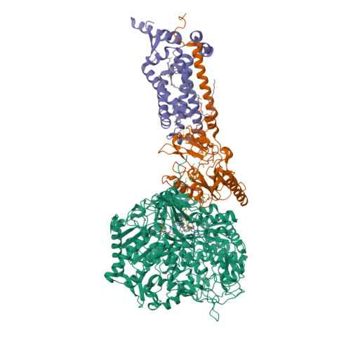
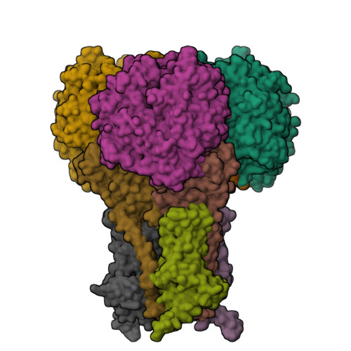
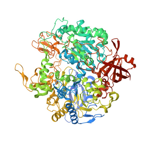
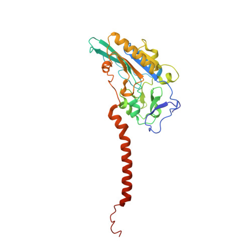
Molecular basis of proton motive force generation: structure of formate dehydrogenase-N.

Jormakka, M.Tornroth, S.Byrne, B.Iwata, S.

(2002) Science 295: 1863-1868

  • DOI: https://doi.org/10.1126/science.1068186
  • Primary Citation of Related Structures:  
    1KQF, 1KQG

  • PubMed Abstract: 

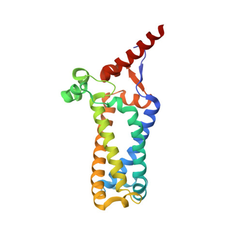
    The structure of the membrane protein formate dehydrogenase-N (Fdn-N), a major component of Escherichia coli nitrate respiration, has been determined at 1.6 angstroms. The structure demonstrates 11 redox centers, including molybdopterin-guanine dinucleotides, five [4Fe-4S] clusters, two heme b groups, and a menaquinone analog. These redox centers are aligned in a single chain, which extends almost 90 angstroms through the enzyme. The menaquinone reduction site associated with a possible proton pathway was also characterized. This structure provides critical insights into the proton motive force generation by redox loop, a common mechanism among a wide range of respiratory enzymes.


  • Organizational Affiliation

    Division of Biomedical Sciences, Imperial College, London SW7 2AZ, UK.


Macromolecules
Find similar proteins by:  (by identity cutoff)  |  3D Structure
Entity ID: 1
MoleculeChains Sequence LengthOrganismDetailsImage
FORMATE DEHYDROGENASE, NITRATE-INDUCIBLE, MAJOR SUBUNIT1,015Escherichia coliMutation(s): 0 
EC: 1.2.1.2 (PDB Primary Data), 1.17.5.3 (UniProt)
Membrane Entity: Yes 
UniProt
Find proteins for P24183 (Escherichia coli (strain K12))
Explore P24183 
Go to UniProtKB:  P24183
Entity Groups  
Sequence Clusters30% Identity50% Identity70% Identity90% Identity95% Identity100% Identity
UniProt GroupP24183
Sequence Annotations
Expand
  • Reference Sequence
Find similar proteins by:  (by identity cutoff)  |  3D Structure
Entity ID: 2
MoleculeChains Sequence LengthOrganismDetailsImage
FORMATE DEHYDROGENASE, NITRATE-INDUCIBLE, IRON-SULFUR SUBUNIT294Escherichia coliMutation(s): 0 
EC: 1.2.1.2
Membrane Entity: Yes 
UniProt
Find proteins for P0AAJ3 (Escherichia coli (strain K12))
Explore P0AAJ3 
Go to UniProtKB:  P0AAJ3
Entity Groups  
Sequence Clusters30% Identity50% Identity70% Identity90% Identity95% Identity100% Identity
UniProt GroupP0AAJ3
Sequence Annotations
Expand
  • Reference Sequence
Find similar proteins by:  (by identity cutoff)  |  3D Structure
Entity ID: 3
MoleculeChains Sequence LengthOrganismDetailsImage
FORMATE DEHYDROGENASE, NITRATE-INDUCIBLE, CYTOCHROME B556(FDN) SUBUNIT217Escherichia coliMutation(s): 0 
EC: 1.2.1.2
Membrane Entity: Yes 
UniProt
Find proteins for P0AEK7 (Escherichia coli (strain K12))
Explore P0AEK7 
Go to UniProtKB:  P0AEK7
Entity Groups  
Sequence Clusters30% Identity50% Identity70% Identity90% Identity95% Identity100% Identity
UniProt GroupP0AEK7
Sequence Annotations
Expand
  • Reference Sequence
Small Molecules
Ligands 5 Unique
IDChains Name / Formula / InChI Key2D Diagram3D Interactions
CDL
Query on CDL

Download Ideal Coordinates CCD File 
N [auth C]CARDIOLIPIN
C81 H156 O17 P2
XVTUQDWPJJBEHJ-KZCWQMDCSA-L
MGD
Query on MGD

Download Ideal Coordinates CCD File 
F [auth A],
G [auth A]
2-AMINO-5,6-DIMERCAPTO-7-METHYL-3,7,8A,9-TETRAHYDRO-8-OXA-1,3,9,10-TETRAAZA-ANTHRACEN-4-ONE GUANOSINE DINUCLEOTIDE
C20 H26 N10 O13 P2 S2
VQAGYJCYOLHZDH-ILXWUORBSA-N
HEM
Query on HEM

Download Ideal Coordinates CCD File 
L [auth C],
M [auth C]
PROTOPORPHYRIN IX CONTAINING FE
C34 H32 Fe N4 O4
KABFMIBPWCXCRK-RGGAHWMASA-L
SF4
Query on SF4

Download Ideal Coordinates CCD File 
E [auth A],
H [auth B],
I [auth B],
J [auth B],
K [auth B]
IRON/SULFUR CLUSTER
Fe4 S4
LJBDFODJNLIPKO-UHFFFAOYSA-N
6MO
Query on 6MO

Download Ideal Coordinates CCD File 
D [auth A]MOLYBDENUM(VI) ION
Mo
HCNGUXXTNNIKCQ-UHFFFAOYSA-N
Experimental Data & Validation

Experimental Data

  • Method: X-RAY DIFFRACTION
  • Resolution: 1.60 Å
  • R-Value Free:  0.195 (Depositor), 0.200 (DCC) 
  • R-Value Work:  0.177 (Depositor), 0.180 (DCC) 
  • R-Value Observed: 0.177 (Depositor) 
Space Group: P 21 3
Unit Cell:
Length ( Å )Angle ( ˚ )
a = 203α = 90
b = 203β = 90
c = 203γ = 90
Software Package:
Software NamePurpose
MLPHAREphasing
CNSrefinement
DENZOdata reduction
SCALEPACKdata scaling

Structure Validation

View Full Validation Report


Ligand Structure Quality Assessment 

Created with Raphaël 2.3.0Worse 01 BetterLigand structure goodness of fit to experimental dataBest fitted CDLClick on this verticalbar to view detailsBest fitted MGDClick on this verticalbar to view detailsBest fitted HEMClick on this verticalbar to view details

Entry History 

Deposition Data

Revision History  (Full details and data files)

  • Version 1.0: 2002-03-15
    Type: Initial release
  • Version 1.1: 2008-04-27
    Changes: Version format compliance
  • Version 1.2: 2011-07-13
    Changes: Derived calculations, Version format compliance
  • Version 1.3: 2014-02-19
    Changes: Atomic model, Structure summary
  • Version 1.4: 2014-08-06
    Changes: Derived calculations, Structure summary
  • Version 1.5: 2024-02-14
    Changes: Data collection, Database references, Derived calculations