Crystal structures of the human p56lck SH2 domain in complex with two short phosphotyrosyl peptides at 1.0 A and 1.8 A resolution.
Tong, L., Warren, T.C., King, J., Betageri, R., Rose, J., Jakes, S.(1996) J Mol Biol 256: 601-610
- PubMed: 8604142
- DOI: https://doi.org/10.1006/jmbi.1996.0112
- Primary Citation of Related Structures:
1LKK, 1LKL - PubMed Abstract:
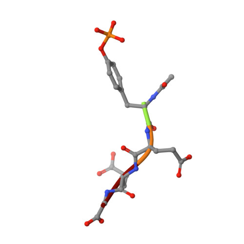
src homology 2 (SH2) domains are modules of about 100 amino acid residues and bind to phosphotyrosine-containing motifs in a sequence-specific manner. They play important roles in intracellular signal transduction and represent potential targets for pharmacological intervention. The protein tyrosine kinase p56lck is a member of the src family and is involved in T-cell activation. The crystal structure of its SH2 domain with an 11-residue peptide showed that the phosphotyrosine and the Ile residue at the pY + 3 position are recognized by the SH2 domain. We present here the crystal structure of the SH2 domain of human p56lck in complex with the short phosphotyrosyl peptide Ac-pTyr-Glu-Glu-Ile (pYEEI peptide) at 1.0 A resolution. The structural analysis at atomic resolution reveals that residue Arg134 (alphaA2), which interacts with the phosphotyrosine side-chain, is present in two conformations in the complex. The structure at 1.8 A resolution of the complex with the phosphotyrosyl peptide Ac-pTyr-Glu-Glu-Gly (pYEEG peptide), which is 11 fold less potent, shows another binding mode for the pY + 3 residue as well as rearrangements of the side-chain of Arg196 (EF3) and one of the water molecules at the base of the pY + 3 pocket. The structure of the complex with the short pYEEI peptide at atomic resolution represents a good starting point for the design and optimization of new inhibitors. Comparative structural analysis of many different inhibitor complexes will be an important component of this drug discovery process.
Organizational Affiliation:
Department of Inflammatory Diseases, Boehringer Ingelheim Pharmaceuticals Inc. Ridgefield, CT 06877, USA.


