1M00

Rat neuronal NOS heme domain with N-butyl-N'-hydroxyguanidine bound


Experimental Data Snapshot

  • Method: X-RAY DIFFRACTION
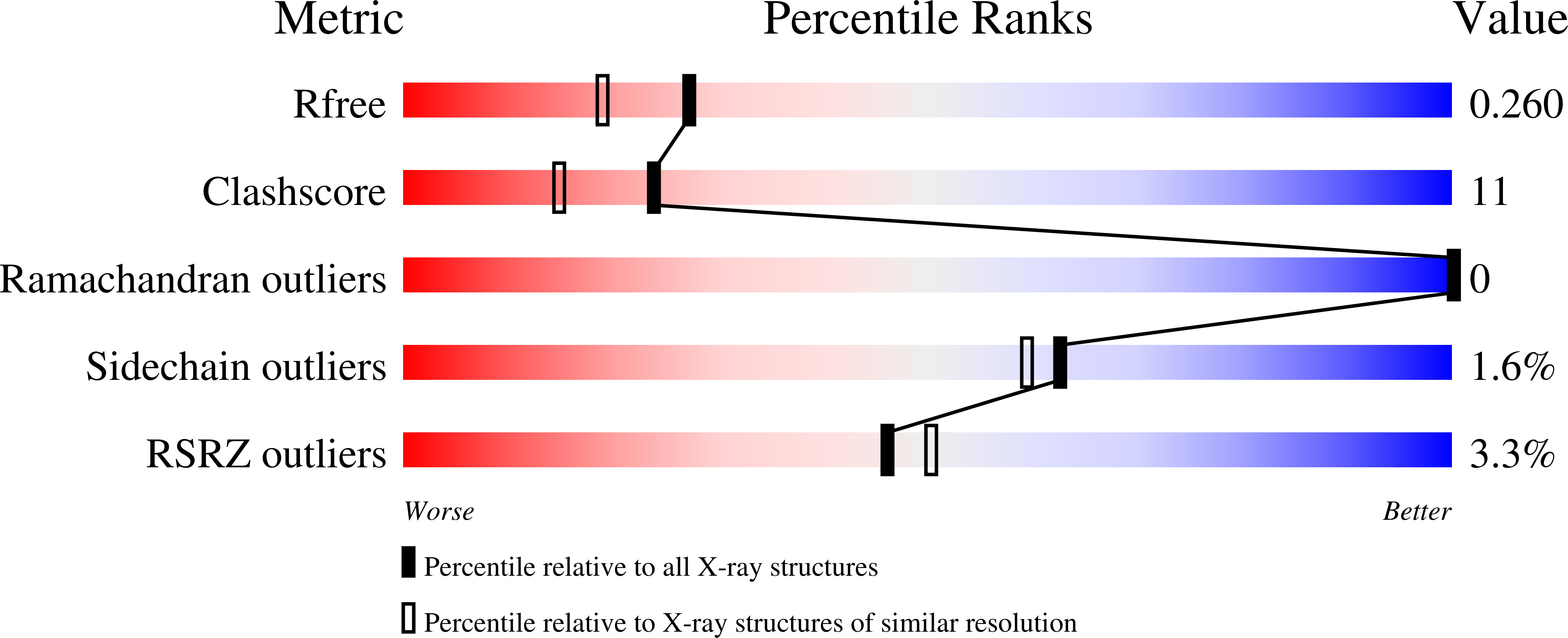
  • Resolution: 2.05 Å
  • R-Value Free: 0.270 
  • R-Value Work: 0.229 

wwPDB Validation   3D Report Full Report


Ligand Structure Quality Assessment 


This is version 1.3 of the entry. See complete history


Literature

The Novel Binding Mode of N-Alkyl-N'-Hydroxyguanidine to Neuronal Nitric Oxide Synthase Provides Mechanistic Insights into NO Biosynthesis

Li, H.Shimizu, H.Flinspach, M.Jamal, J.Yang, W.Xian, M.Cai, T.Wen, E.Z.Jia, Q.Wang, P.G.Poulos, T.L.

(2002) Biochemistry 41: 13868-13875

  • DOI: https://doi.org/10.1021/bi020417c
  • Primary Citation of Related Structures:  
    1LZX, 1LZZ, 1M00

  • PubMed Abstract: 

    A series of N-alkyl-N'-hydroxyguanidine compounds have recently been characterized as non-amino acid substrates for all three nitric oxide synthase (NOS) isoforms which mimic NO formation from N(omega)-hydroxy-L-arginine. Crystal structures of the nNOS heme domain complexed with either N-isopropyl-N'-hydroxyguanidine or N-butyl-N'-hydroxyguanidine reveal two different binding modes in the substrate binding pocket. The binding mode of the latter is consistent with that observed for the substrate N(omega)-hydroxy-L-arginine bound in the nNOS active site. However, the former binds to nNOS in an unexpected fashion, thus providing new insights into the mechanism on how the hydroxyguanidine moiety leads to NO formation. Structural features of substrate binding support the view that the OH-substituted guanidine nitrogen, instead of the hydroxyl oxygen, is the source of hydrogen supplied to the active ferric-superoxy species for the second step of the NOS catalytic reaction.


  • Organizational Affiliation

    Department of Molecular Biology and Biochemistry and Program in Macromolecular Structures, University of California, Irvine, California 92697, USA.


Macromolecules
Find similar proteins by:  (by identity cutoff)  |  3D Structure
Entity ID: 1
MoleculeChains Sequence LengthOrganismDetailsImage
Nitric-oxide synthase
A, B
419Rattus norvegicusMutation(s): 0 
EC: 1.14.13.39
UniProt
Find proteins for P29476 (Rattus norvegicus)
Explore P29476 
Go to UniProtKB:  P29476
Entity Groups  
Sequence Clusters30% Identity50% Identity70% Identity90% Identity95% Identity100% Identity
UniProt GroupP29476
Sequence Annotations
Expand
  • Reference Sequence
Small Molecules
Ligands 5 Unique
IDChains Name / Formula / InChI Key2D Diagram3D Interactions
HEM
Query on HEM

Download Ideal Coordinates CCD File 
E [auth A],
I [auth B]
PROTOPORPHYRIN IX CONTAINING FE
C34 H32 Fe N4 O4
KABFMIBPWCXCRK-RGGAHWMASA-L
H4B
Query on H4B

Download Ideal Coordinates CCD File 
F [auth A],
J [auth B]
5,6,7,8-TETRAHYDROBIOPTERIN
C9 H15 N5 O3
FNKQXYHWGSIFBK-RPDRRWSUSA-N
BHH
Query on BHH

Download Ideal Coordinates CCD File 
G [auth A],
K [auth B]
N-BUTYL-N'-HYDROXYGUANIDINE
C5 H13 N3 O
ULDDTFAPYWLDGF-UHFFFAOYSA-N
ZN
Query on ZN

Download Ideal Coordinates CCD File 
D [auth A]ZINC ION
Zn
PTFCDOFLOPIGGS-UHFFFAOYSA-N
ACT
Query on ACT

Download Ideal Coordinates CCD File 
C [auth A],
H [auth B]
ACETATE ION
C2 H3 O2
QTBSBXVTEAMEQO-UHFFFAOYSA-M
Experimental Data & Validation

Experimental Data

  • Method: X-RAY DIFFRACTION
  • Resolution: 2.05 Å
  • R-Value Free: 0.270 
  • R-Value Work: 0.229 
  • Space Group: P 21 21 21
Unit Cell:
Length ( Å )Angle ( ˚ )
a = 52.07α = 90
b = 111.25β = 90
c = 163.84γ = 90
Software Package:
Software NamePurpose
DENZOdata reduction
SCALEPACKdata scaling
CNSrefinement
CNSphasing

Structure Validation

View Full Validation Report


Ligand Structure Quality Assessment 


Entry History 

Deposition Data

Revision History  (Full details and data files)

  • Version 1.0: 2002-11-27
    Type: Initial release
  • Version 1.1: 2008-04-28
    Changes: Version format compliance
  • Version 1.2: 2011-07-13
    Changes: Version format compliance
  • Version 1.3: 2024-02-14
    Changes: Data collection, Database references, Derived calculations