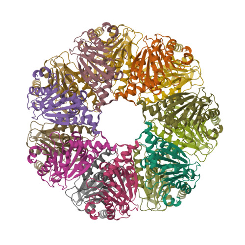
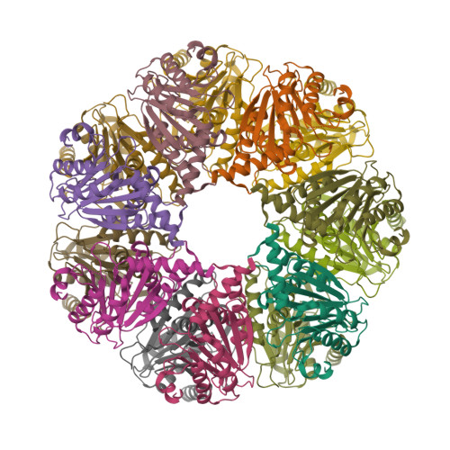
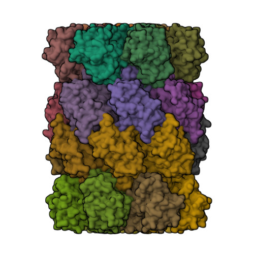
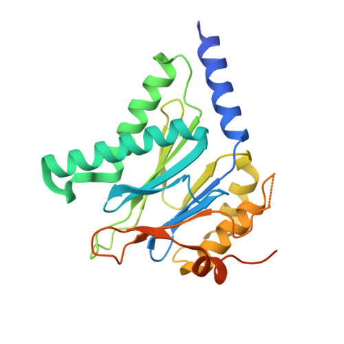
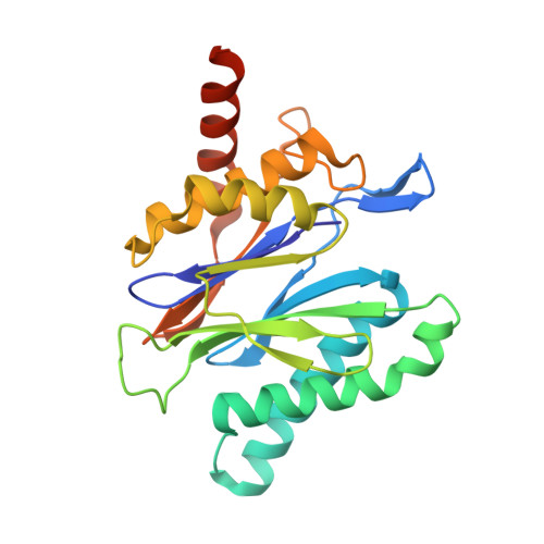
1Q5Q

The Rhodococcus 20S proteasome


Experimental Data Snapshot

  • Method: X-RAY DIFFRACTION
  • Resolution: 2.60 Å
  • R-Value Free: 0.264 
  • R-Value Work: 0.246 
  • R-Value Observed: 0.246 

Starting Model: experimental
View more details

wwPDB Validation   3D Report Full Report


This is version 1.3 of the entry. See complete history


Literature

Crystal structures of the Rhodococcus proteasome with and without its pro-peptides: implications for the role of the pro-peptide in proteasome assembly.

Kwon, Y.D.Nagy, I.Adams, P.D.Baumeister, W.Jap, B.K.

(2004) J Mol Biol 335: 233-245

  • DOI: https://doi.org/10.1016/j.jmb.2003.08.029
  • Primary Citation of Related Structures:  
    1Q5Q, 1Q5R

  • PubMed Abstract: 

    To understand the role of the pro-peptide in proteasome assembly, we have determined structures of the Rhodococcus proteasome and a mutant form that prevents the autocatalytic removal of its pro-peptides. The structures reveal that the pro-peptide acts as an assembly-promoting factor by linking its own beta-subunit to two adjacent alpha-subunits, thereby providing a molecular explanation for the observed kinetics of proteasome assembly. The Rhodococcus proteasome has been found to have a substantially smaller contact region between alpha-subunits compared to those regions in the proteasomes of Thermoplasma, yeast, and mammalian cells, suggesting that a smaller contact area between alpha-subunits is likely the structural basis for the Rhodococcus alpha-subunits not assembling into alpha-rings when expressed alone. Analysis of all available beta-subunit structures shows that the contact area between beta-subunits within a beta-ring is not sufficient for beta-ring self-assembly without the additional contact provided by the alpha-ring. This appears to be a fail-safe mechanism ensuring that the active sites on the beta-subunits are activated only after proteasome assembly is complete.


  • Organizational Affiliation

    Graduate Group in Comparative Biochemistry, University of California, Berkeley, CA 94720, USA.


Macromolecules
Find similar proteins by:  (by identity cutoff)  |  3D Structure
Entity ID: 1
MoleculeChains Sequence LengthOrganismDetailsImage
proteasome alpha-type subunit 1
A, B, C, D, E
A, B, C, D, E, F, G
259Rhodococcus erythropolisMutation(s): 0 
Gene Names: PRCA(1)
EC: 3.4.25.1
UniProt
Find proteins for Q53080 (Rhodococcus erythropolis)
Explore Q53080 
Go to UniProtKB:  Q53080
Entity Groups  
Sequence Clusters30% Identity50% Identity70% Identity90% Identity95% Identity100% Identity
UniProt GroupQ53080
Sequence Annotations
Expand
  • Reference Sequence
Find similar proteins by:  (by identity cutoff)  |  3D Structure
Entity ID: 2
MoleculeChains Sequence LengthOrganismDetailsImage
proteasome beta-type subunit 1
H, I, J, K, L
H, I, J, K, L, M, N
235Rhodococcus erythropolisMutation(s): 0 
Gene Names: PRCB(1)
EC: 3.4.25.1
UniProt
Find proteins for Q53079 (Rhodococcus erythropolis)
Explore Q53079 
Go to UniProtKB:  Q53079
Entity Groups  
Sequence Clusters30% Identity50% Identity70% Identity90% Identity95% Identity100% Identity
UniProt GroupQ53079
Sequence Annotations
Expand
  • Reference Sequence
Experimental Data & Validation

Experimental Data

  • Method: X-RAY DIFFRACTION
  • Resolution: 2.60 Å
  • R-Value Free: 0.264 
  • R-Value Work: 0.246 
  • R-Value Observed: 0.246 
  • Space Group: C 2 2 21
Unit Cell:
Length ( Å )Angle ( ˚ )
a = 150.112α = 90
b = 215.307β = 90
c = 251.051γ = 90
Software Package:
Software NamePurpose
CNSrefinement
DENZOdata reduction
SCALEPACKdata scaling
CNSphasing

Structure Validation

View Full Validation Report


Entry History 

Deposition Data

Revision History  (Full details and data files)

  • Version 1.0: 2003-12-16
    Type: Initial release
  • Version 1.1: 2008-04-29
    Changes: Version format compliance
  • Version 1.2: 2011-07-13
    Changes: Derived calculations, Version format compliance
  • Version 1.3: 2023-08-16
    Changes: Data collection, Database references, Refinement description