1QZF

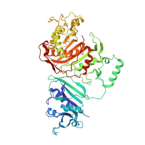
Crystal structure of DHFR-TS from Cryptosporidium hominis


Experimental Data Snapshot

  • Method: X-RAY DIFFRACTION
  • Resolution: 2.80 Å
  • R-Value Free: 0.245 
  • R-Value Work: 0.225 
  • R-Value Observed: 0.225 

Starting Models: experimental
View more details

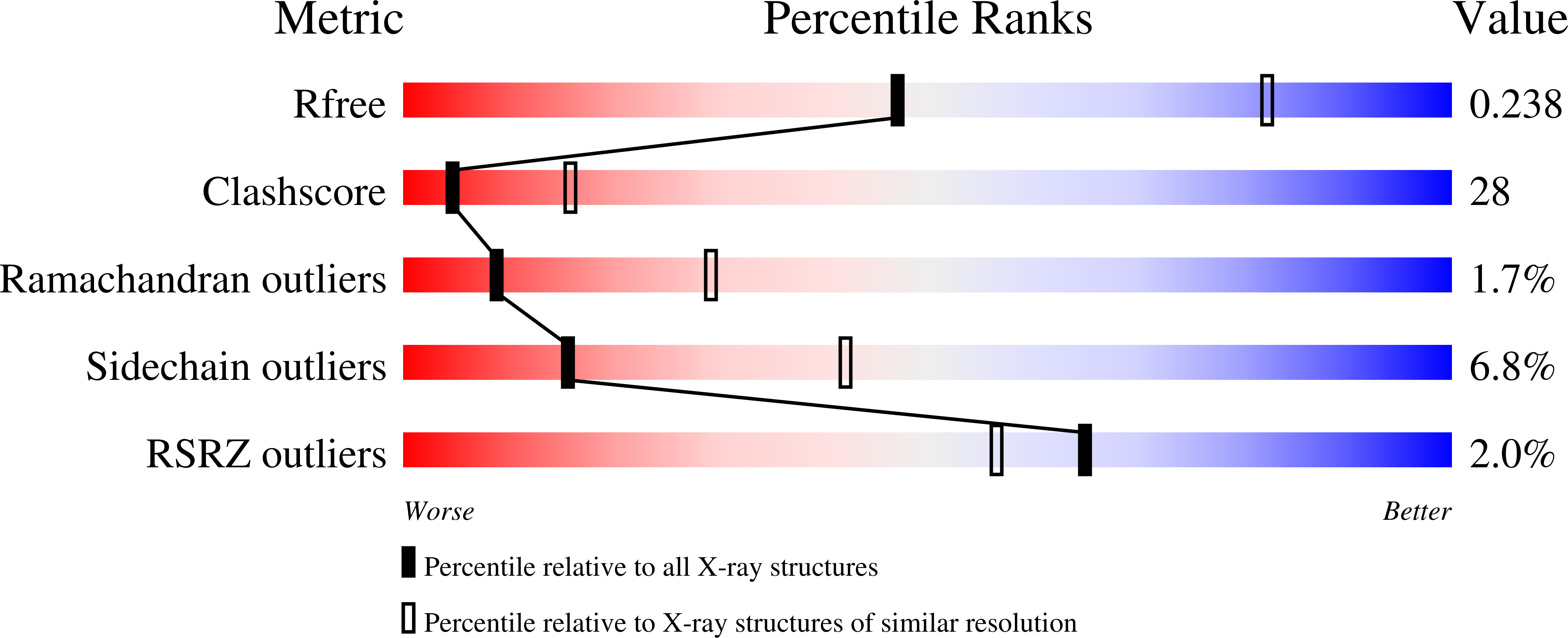
wwPDB Validation   3D Report Full Report


Ligand Structure Quality Assessment 


This is version 1.3 of the entry. See complete history


Literature

Phylogenetic classification of protozoa based on the structure of the linker domain in the bifunctional enzyme, dihydrofolate reductase-thymidylate synthase

O'Neil, R.H.Lilien, R.H.Donald, B.R.Stroud, R.M.Anderson, A.C.

(2003) J Biol Chem 278: 52980-52987

  • DOI: https://doi.org/10.1074/jbc.M310328200
  • Primary Citation of Related Structures:  
    1QZF

  • PubMed Abstract: 

    We have determined the crystal structure of dihydrofolate reductase-thymidylate synthase (DHFR-TS) from Cryptosporidium hominis, revealing a unique linker domain containing an 11-residue alpha-helix that has extensive interactions with the opposite DHFR-TS monomer of the homodimeric enzyme. Analysis of the structure of DHFR-TS from C. hominis and of previously solved structures of DHFR-TS from Plasmodium falciparum and Leishmania major reveals that the linker domain primarily controls the relative orientation of the DHFR and TS domains. Using the tertiary structure of the linker domains, we have been able to place a number of protozoa in two distinct and dissimilar structural families corresponding to two evolutionary families and provide the first structural evidence validating the use of DHFR-TS as a tool of phylogenetic classification. Furthermore, the structure of C. hominis DHFR-TS calls into question surface electrostatic channeling as the universal means of dihydrofolate transport between TS and DHFR in the bifunctional enzyme.


  • Organizational Affiliation

    Department of Chemistry, Dartmouth College, Hanover, New Hampshire 03755, USA.


Macromolecules
Find similar proteins by:  (by identity cutoff)  |  3D Structure
Entity ID: 1
MoleculeChains Sequence LengthOrganismDetailsImage
bifunctional dihydrofolate reductase-thymidylate synthase
A, B, C, D, E
521Cryptosporidium hominisMutation(s): 0 
EC: 1.5.1.3 (PDB Primary Data), 2.1.1.45 (PDB Primary Data)
UniProt
Find proteins for Q27552 (Cryptosporidium parvum)
Explore Q27552 
Go to UniProtKB:  Q27552
Entity Groups  
Sequence Clusters30% Identity50% Identity70% Identity90% Identity95% Identity100% Identity
UniProt GroupQ27552
Sequence Annotations
Expand
  • Reference Sequence
Small Molecules
Ligands 4 Unique
IDChains Name / Formula / InChI Key2D Diagram3D Interactions
NDP
Query on NDP

Download Ideal Coordinates CCD File 
I [auth A],
M [auth B],
Q [auth C],
U [auth D],
Y [auth E]
NADPH DIHYDRO-NICOTINAMIDE-ADENINE-DINUCLEOTIDE PHOSPHATE
C21 H30 N7 O17 P3
ACFIXJIJDZMPPO-NNYOXOHSSA-N
CB3
Query on CB3

Download Ideal Coordinates CCD File 
G [auth A],
K [auth B],
O [auth C],
S [auth D],
W [auth E]
10-PROPARGYL-5,8-DIDEAZAFOLIC ACID
C24 H23 N5 O6
LTKHPMDRMUCUEB-IBGZPJMESA-N
FOL
Query on FOL

Download Ideal Coordinates CCD File 
H [auth A],
L [auth B],
P [auth C],
T [auth D],
X [auth E]
FOLIC ACID
C19 H19 N7 O6
OVBPIULPVIDEAO-LBPRGKRZSA-N
UMP
Query on UMP

Download Ideal Coordinates CCD File 
F [auth A],
J [auth B],
N [auth C],
R [auth D],
V [auth E]
2'-DEOXYURIDINE 5'-MONOPHOSPHATE
C9 H13 N2 O8 P
JSRLJPSBLDHEIO-SHYZEUOFSA-N
Experimental Data & Validation

Experimental Data

  • Method: X-RAY DIFFRACTION
  • Resolution: 2.80 Å
  • R-Value Free: 0.245 
  • R-Value Work: 0.225 
  • R-Value Observed: 0.225 
  • Space Group: C 1 2 1
Unit Cell:
Length ( Å )Angle ( ˚ )
a = 214.9α = 90
b = 116.3β = 95.23
c = 219.7γ = 90
Software Package:
Software NamePurpose
CNSrefinement
DENZOdata reduction
SCALEPACKdata scaling
CNSphasing

Structure Validation

View Full Validation Report


Ligand Structure Quality Assessment 


Entry History 

Deposition Data

Revision History  (Full details and data files)

  • Version 1.0: 2003-11-11
    Type: Initial release
  • Version 1.1: 2008-04-29
    Changes: Version format compliance
  • Version 1.2: 2011-07-13
    Changes: Version format compliance
  • Version 1.3: 2023-08-23
    Changes: Data collection, Database references, Derived calculations, Refinement description