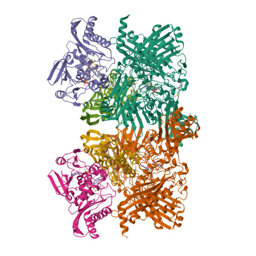
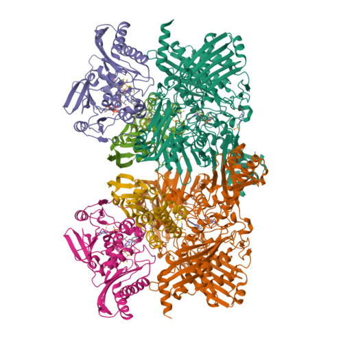
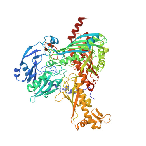
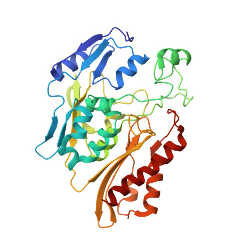
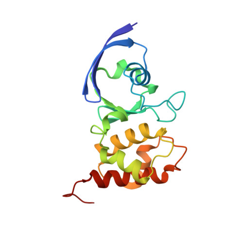
1SB3

Structure of 4-hydroxybenzoyl-CoA reductase from Thauera aromatica


Experimental Data Snapshot

  • Method: X-RAY DIFFRACTION
  • Resolution: 2.20 Å
  • R-Value Free: 0.205 
  • R-Value Work: 0.171 
  • R-Value Observed: 0.171 

Starting Model: experimental
View more details

wwPDB Validation   3D Report Full Report


Ligand Structure Quality Assessment 


This is version 1.3 of the entry. See complete history


Literature

Structure of a Xanthine Oxidase-Related 4-Hydroxybenzoyl-CoA Reductase with an Additional [4Fe-4S] Cluster and an Inverted Electron Flow.

Unciuleac, M.Warkentin, E.Page, C.C.Boll, M.Ermler, U.

(2004) Structure 12: 2249-2256

  • DOI: https://doi.org/10.1016/j.str.2004.10.008
  • Primary Citation of Related Structures:  
    1RM6, 1SB3

  • PubMed Abstract: 

    The Mo-flavo-Fe/S-dependent heterohexameric protein complex 4-hydroxybenzoyl-CoA reductase (4-HBCR, dehydroxylating) is a central enzyme of the anaerobic degradation of phenolic compounds and belongs to the xanthine oxidase (XO) family of molybdenum enzymes. Its X-ray structure was established at 1.6 A resolution. The most pronounced difference between 4-HBCR and other structurally characterized members of the XO family is the insertion of 40 amino acids within the beta subunit, which carries an additional [4Fe-4S] cluster at a distance of 16.5 A to the isoalloxazine ring of FAD. The architecture of 4-HBCR and concomitantly performed electron transfer rate calculations suggest an inverted electron transfer chain from the donor ferredoxin via the [4Fe-4S] cluster to the Mo over a distance of 55 A. The binding site of 4-hydroxybenzoyl-CoA is located in an 18 A long channel lined up by several aromatic side chains around the aromatic moiety, which are proposed to shield and stabilize the postulated radical intermediates during catalysis.


  • Organizational Affiliation

    Institut für Biologie II, Mikrobiologie Schänzlestrasse 1, D-79104 Freiburg, Germany.


Macromolecules
Find similar proteins by:  (by identity cutoff)  |  3D Structure
Entity ID: 1
MoleculeChains Sequence LengthOrganismDetailsImage
4-hydroxybenzoyl-CoA reductase alpha subunit
A, D
769Thauera aromaticaMutation(s): 0 
EC: 1.3.99.20 (PDB Primary Data), 1.1.7.1 (UniProt)
UniProt
Find proteins for O33819 (Thauera aromatica)
Explore O33819 
Go to UniProtKB:  O33819
Entity Groups  
Sequence Clusters30% Identity50% Identity70% Identity90% Identity95% Identity100% Identity
UniProt GroupO33819
Sequence Annotations
Expand
  • Reference Sequence
Find similar proteins by:  (by identity cutoff)  |  3D Structure
Entity ID: 2
MoleculeChains Sequence LengthOrganismDetailsImage
4-hydroxybenzoyl-CoA reductase beta subunit
B, E
324Thauera aromaticaMutation(s): 0 
EC: 1.3.99.20 (PDB Primary Data), 1.1.7.1 (UniProt)
UniProt
Find proteins for O33820 (Thauera aromatica)
Explore O33820 
Go to UniProtKB:  O33820
Entity Groups  
Sequence Clusters30% Identity50% Identity70% Identity90% Identity95% Identity100% Identity
UniProt GroupO33820
Sequence Annotations
Expand
  • Reference Sequence
Find similar proteins by:  (by identity cutoff)  |  3D Structure
Entity ID: 3
MoleculeChains Sequence LengthOrganismDetailsImage
4-hydroxybenzoyl-CoA reductase gamma subunit
C, F
161Thauera aromaticaMutation(s): 0 
EC: 1.3.99.20 (PDB Primary Data), 1.1.7.1 (UniProt)
UniProt
Find proteins for O33818 (Thauera aromatica)
Explore O33818 
Go to UniProtKB:  O33818
Entity Groups  
Sequence Clusters30% Identity50% Identity70% Identity90% Identity95% Identity100% Identity
UniProt GroupO33818
Sequence Annotations
Expand
  • Reference Sequence
Small Molecules
Ligands 6 Unique
IDChains Name / Formula / InChI Key2D Diagram3D Interactions
PCD
Query on PCD

Download Ideal Coordinates CCD File 
G [auth A],
N [auth D]
(MOLYBDOPTERIN-CYTOSINE DINUCLEOTIDE-S,S)-DIOXO-AQUA-MOLYBDENUM(V)
C19 H26 Mo N8 O16 P2 S2
YEBYDVFRFUQMER-MQPNXHJTSA-L
FAD
Query on FAD

Download Ideal Coordinates CCD File 
I [auth B],
P [auth E]
FLAVIN-ADENINE DINUCLEOTIDE
C27 H33 N9 O15 P2
VWWQXMAJTJZDQX-UYBVJOGSSA-N
SF4
Query on SF4

Download Ideal Coordinates CCD File 
J [auth B],
Q [auth E]
IRON/SULFUR CLUSTER
Fe4 S4
LJBDFODJNLIPKO-UHFFFAOYSA-N
EPE
Query on EPE

Download Ideal Coordinates CCD File 
H [auth A]4-(2-HYDROXYETHYL)-1-PIPERAZINE ETHANESULFONIC ACID
C8 H18 N2 O4 S
JKMHFZQWWAIEOD-UHFFFAOYSA-N
FES
Query on FES

Download Ideal Coordinates CCD File 
K [auth C],
L [auth C],
R [auth F],
S [auth F]
FE2/S2 (INORGANIC) CLUSTER
Fe2 S2
NIXDOXVAJZFRNF-UHFFFAOYSA-N
SO4
Query on SO4

Download Ideal Coordinates CCD File 
M [auth D],
O [auth E]
SULFATE ION
O4 S
QAOWNCQODCNURD-UHFFFAOYSA-L
Experimental Data & Validation

Experimental Data

  • Method: X-RAY DIFFRACTION
  • Resolution: 2.20 Å
  • R-Value Free: 0.205 
  • R-Value Work: 0.171 
  • R-Value Observed: 0.171 
  • Space Group: P 21 21 21
Unit Cell:
Length ( Å )Angle ( ˚ )
a = 116.62α = 90
b = 150.2β = 90
c = 175.27γ = 90
Software Package:
Software NamePurpose
CNSrefinement
DENZOdata reduction
SCALEPACKdata scaling
EPMRphasing

Structure Validation

View Full Validation Report


Ligand Structure Quality Assessment 


Entry History 

Deposition Data

Revision History  (Full details and data files)

  • Version 1.0: 2004-12-21
    Type: Initial release
  • Version 1.1: 2008-04-29
    Changes: Version format compliance
  • Version 1.2: 2011-07-13
    Changes: Version format compliance
  • Version 1.3: 2023-08-23
    Changes: Data collection, Database references, Derived calculations, Refinement description