Mapping Protein Matrix Cavities in Human Cytoglobin Through Xe Atom Binding
De Sanctis, D., Dewilde, S., Pesce, A., Moens, L., Ascenzi, P., Hankeln, T., Burmester, T., Bolognesi, M.(2004) Biochem Biophys Res Commun 316: 1217
- PubMed: 15044115
- DOI: https://doi.org/10.1016/j.bbrc.2004.03.007
- Primary Citation of Related Structures:
1URY, 1UX9 - PubMed Abstract:
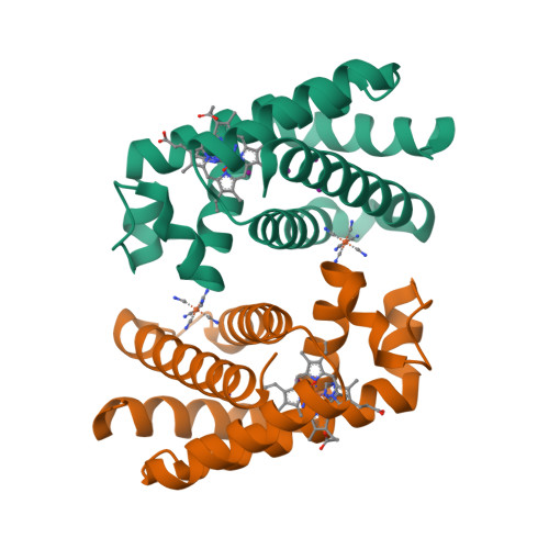
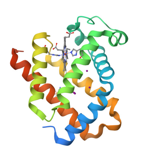
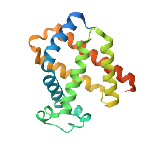
Cytoglobin is the fourth recognized globin type, almost ubiquitously distributed in human tissues; its function is still poorly understood. Cytoglobin displays a core region of about 150 residues, structurally related to hemoglobin and myoglobin, and two extra segments, about 20 residues each, at the N- and C-termini. The core region hosts a large apolar cavity, held to provide a ligand diffusion pathway to/from the heme, and/or ligand temporary docking sites. Here we report the crystal structure (2.4A resolution, R-factor 19.1%) of a human cytoglobin mutant bearing the CysB2(38) --> Ser and CysE9(83) --> Ser substitutions (CYGB*), treated under pressurized xenon. Three Xe atoms bind to the heme distal site region of CYGB* mapping the protein matrix apolar cavity. Despite the conserved globin fold, the cavity found in CYGB* is structured differently from those recognized to play a functional role in myoglobin, neuroglobin, truncated hemoglobins, and Cerebratulus lacteus mini-hemoglobin.
Organizational Affiliation:
Department of Physics-INFM and Centre for Excellence in Biomedical Research, University of Genova, Via Dodecaneso 33, Genoa I-16146, Italy.


