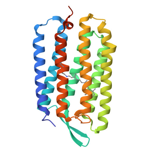
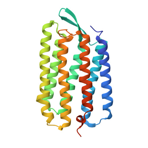
Deformation of helix C in the low temperature L-intermediate of bacteriorhodopsin.
Edman, K., Royant, A., Larsson, G., Jacobson, F., Taylor, T., van der Spoel, D., Landau, E.M., Pebay-Peyroula, E., Neutze, R.(2004) J Biological Chem 279: 2147-2158
- PubMed: 14532280
- DOI: https://doi.org/10.1074/jbc.M300709200
- Primary Citation of Related Structures:
1VJM - PubMed Abstract:
X-ray and electron diffraction studies of specific reaction intermediates, or reaction intermediate analogues, have produced a consistent picture of the structural mechanism of light-driven proton pumping by bacteriorhodopsin. Of central importance within this picture is the structure of the L-intermediate, which follows the retinal all-trans to 13-cis photoisomerization step of the K-intermediate and sets the stage for the primary proton transfer event from the positively charged Schiff base to the negatively charged Asp-85. Here we report the structural changes in bacteriorhodopsin following red light illumination at 150 K. Single crystal microspectrophotometry showed that only the L-intermediate is populated in three-dimensional crystals under these conditions. The experimental difference Fourier electron density map and refined crystallographic structure were consistent with those previously presented (Royant, A., Edman, K., Ursby, T., Pebay-Peyroula, E., Landau, E. M., and Neutze, R. (2000) Nature 406, 645-648; Royant, A., Edman, K., Ursby, T., Pebay-Peyroula, E., Landau, E. M., and Neutze, R. (2001) Photochem. Photobiol. 74, 794-804). Based on the refined crystallographic structures, molecular dynamic simulations were used to examine the influence of the conformational change of the protein that is associated with the K-to-L transition on retinal dynamics. Implications regarding the structural mechanism for proton pumping by bacteriorhodopsin are discussed.
Organizational Affiliation:
Department of Chemistry and Bioscience, Chalmers University of Technology, Box 462, S-40530 Gothenburg, Sweden.