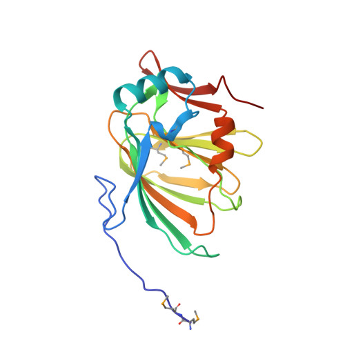
1X82

CRYSTAL STRUCTURE OF PHOSPHOGLUCOSE ISOMERASE FROM PYROCOCCUS FURIOSUS WITH BOUND 5-phospho-D-arabinonate


Experimental Data Snapshot

  • Method: X-RAY DIFFRACTION
  • Resolution: 1.50 Å
  • R-Value Free: 0.208 
  • R-Value Work: 0.161 
  • R-Value Observed: 0.163 

Starting Model: experimental
View more details

wwPDB Validation   3D Report Full Report


Ligand Structure Quality Assessment 


This is version 1.6 of the entry. See complete history


Literature

The structures of inhibitor complexes of Pyrococcus furiosus phosphoglucose isomerase provide insights into substrate binding and catalysis.

Berrisford, J.M.Akerboom, J.Brouns, S.Sedelnikova, S.E.Turnbull, A.P.van der Oost, J.Salmon, L.Hardre, R.Murray, I.A.Blackburn, G.M.Rice, D.W.Baker, P.J.

(2004) J Mol Biol 343: 649-657

  • DOI: https://doi.org/10.1016/j.jmb.2004.08.061
  • Primary Citation of Related Structures:  
    1X7N, 1X82, 1X8E

  • PubMed Abstract: 

    Pyrococcus furiosus phosphoglucose isomerase (PfPGI) is a metal-containing enzyme that catalyses the interconversion of glucose 6-phosphate (G6P) and fructose 6-phosphate (F6P). The recent structure of PfPGI has confirmed the hypothesis that the enzyme belongs to the cupin superfamily and identified the position of the active site. This fold is distinct from the alphabetaalpha sandwich fold commonly seen in phosphoglucose isomerases (PGIs) that are found in bacteria, eukaryotes and some archaea. Whilst the mechanism of the latter family is thought to proceed through a cis-enediol intermediate, analysis of the structure of PfPGI in the presence of inhibitors has led to the suggestion that the mechanism of this enzyme involves the metal-dependent direct transfer of a hydride between C1 and C2 atoms of the substrate. To gain further insight in the reaction mechanism of PfPGI, the structures of the free enzyme and the complexes with the inhibitor, 5-phospho-d-arabinonate (5PAA) in the presence and absence of metal have been determined. Comparison of these structures with those of equivalent complexes of the eukaryotic PGIs reveals similarities at the active site in the disposition of possible catalytic residues. These include the presence of a glutamic acid residue, Glu97 in PfPGI, which occupies the same position relative to the inhibitor as that of the glutamate that is thought to function as the catalytic base in the eukaryal-type PGIs. These similarities suggest that aspects of the catalytic mechanisms of these two structurally unrelated PGIs may be similar and based on an enediol intermediate.


  • Organizational Affiliation

    The Krebs Institute for Biomolecular Research, Department of Molecular Biology and Biotechnology, University of Sheffield, Firth Court, Western Bank, Sheffield S10 2TN, UK.


Macromolecules
Find similar proteins by:  (by identity cutoff)  |  3D Structure
Entity ID: 1
MoleculeChains Sequence LengthOrganismDetailsImage
Glucose-6-phosphate isomerase190Pyrococcus furiosusMutation(s): 4 
Gene Names: pgiA
EC: 5.3.1.9
UniProt
Find proteins for P83194 (Pyrococcus furiosus (strain ATCC 43587 / DSM 3638 / JCM 8422 / Vc1))
Explore P83194 
Go to UniProtKB:  P83194
Entity Groups  
Sequence Clusters30% Identity50% Identity70% Identity90% Identity95% Identity100% Identity
UniProt GroupP83194
Sequence Annotations
Expand
  • Reference Sequence
Small Molecules
Ligands 1 Unique
IDChains Name / Formula / InChI Key2D Diagram3D Interactions
PA5
Query on PA5

Download Ideal Coordinates CCD File 
B [auth A]5-PHOSPHOARABINONIC ACID
C5 H11 O9 P
HNECGPFIYSOYHF-JJYYJPOSSA-N
Modified Residues  1 Unique
IDChains TypeFormula2D DiagramParent
MSE
Query on MSE
A
L-PEPTIDE LINKINGC5 H11 N O2 SeMET
Experimental Data & Validation

Experimental Data

  • Method: X-RAY DIFFRACTION
  • Resolution: 1.50 Å
  • R-Value Free: 0.208 
  • R-Value Work: 0.161 
  • R-Value Observed: 0.163 
  • Space Group: C 1 2 1
Unit Cell:
Length ( Å )Angle ( ˚ )
a = 84.506α = 90
b = 42.73β = 120.32
c = 57.308γ = 90
Software Package:
Software NamePurpose
DENZOdata reduction
SCALEPACKdata scaling
REFMACrefinement

Structure Validation

View Full Validation Report


Ligand Structure Quality Assessment 


Entry History 

Deposition Data

Revision History  (Full details and data files)

  • Version 1.0: 2004-10-12
    Type: Initial release
  • Version 1.1: 2008-04-30
    Changes: Version format compliance
  • Version 1.2: 2011-07-13
    Changes: Derived calculations, Version format compliance
  • Version 1.3: 2014-09-24
    Changes: Database references
  • Version 1.4: 2023-08-23
    Changes: Data collection, Database references, Derived calculations, Refinement description
  • Version 1.5: 2023-11-15
    Changes: Data collection
  • Version 1.6: 2024-11-20
    Changes: Structure summary