1YDR

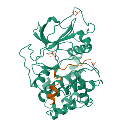
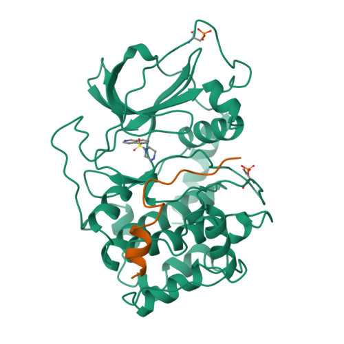
STRUCTURE OF CAMP-DEPENDENT PROTEIN KINASE, ALPHA-CATALYTIC SUBUNIT IN COMPLEX WITH H7 PROTEIN KINASE INHIBITOR 1-(5-ISOQUINOLINESULFONYL)-2-METHYLPIPERAZINE


Experimental Data Snapshot

  • Method: X-RAY DIFFRACTION
  • Resolution: 2.20 Å
  • R-Value Work: 0.188 
  • R-Value Observed: 0.188 

wwPDB Validation   3D Report Full Report


Ligand Structure Quality Assessment 


This is version 1.4 of the entry. See complete history


Literature

Crystal structures of catalytic subunit of cAMP-dependent protein kinase in complex with isoquinolinesulfonyl protein kinase inhibitors H7, H8, and H89. Structural implications for selectivity.

Engh, R.A.Girod, A.Kinzel, V.Huber, R.Bossemeyer, D.

(1996) J Biol Chem 271: 26157-26164

  • DOI: https://doi.org/10.1074/jbc.271.42.26157
  • Primary Citation of Related Structures:  
    1YDR, 1YDS, 1YDT

  • PubMed Abstract: 

    The discovery of several hundred different protein kinases involved in highly diverse cellular signaling pathways is in stark contrast to the much smaller number of known modulators of cell signaling. Of these, the H series protein kinase inhibitors (1-(5-isoquinolinesulfonyl)-2-methylpiperazine (H7), N-[2-(methylamino)ethyl]-5-isoquinolinesulfonamide (H8) N-[2-(p-Bromocinnamylamino)ethyl]-5-isoquinolinesulfonamide (H89)) are frequently used to block signaling pathways in studies of cellular regulation. To elucidate inhibition mechanisms at atomic resolution and to enable structure-based drug design of potential therapeutic modulators of signaling pathways, we determined the crystal structures of corresponding complexes with the cAPK catalytic subunit. Complexes with H7 and H8 (2.2 A) and with H89 (2.3 A) define the binding mode of the isoquinoline-sulfonamide derivatives in the ATP-binding site while demonstrating effects of ligand-induced structural change. Specific interactions between the enzyme and the inhibitors include the isoquinoline ring nitrogen ligating to backbone amide of Val-123 and an inhibitor side chain amide bonding to the backbone carbonyl of Glu-170. The conservation of the ATP-binding site of protein kinases allows evaluation of factors governing general selectivity of these inhibitors among kinases. These results should assist efforts in the design of protein kinase inhibitors with specific properties.


  • Organizational Affiliation

    Abteilung Strukturforschung II, Max-Planck Institute for Biochemistry, D-82152 Martinsried, Federal Republic of Germany.


Macromolecules
Find similar proteins by:  (by identity cutoff)  |  3D Structure
Entity ID: 1
MoleculeChains Sequence LengthOrganismDetailsImage
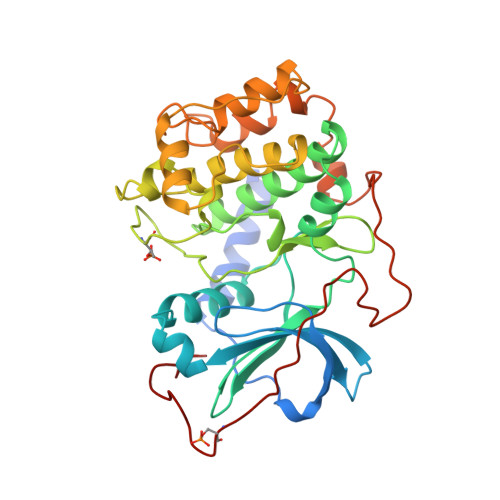
C-AMP-DEPENDENT PROTEIN KINASEA [auth E]350Bos taurusMutation(s): 2 
EC: 2.7.1.37 (PDB Primary Data), 2.7.11.11 (UniProt)
UniProt
Find proteins for P00517 (Bos taurus)
Explore P00517 
Go to UniProtKB:  P00517
Entity Groups  
Sequence Clusters30% Identity50% Identity70% Identity90% Identity95% Identity100% Identity
UniProt GroupP00517
Sequence Annotations
Expand
  • Reference Sequence

Find similar proteins by:  Sequence   |   3D Structure  

Entity ID: 2
MoleculeChains Sequence LengthOrganismDetailsImage
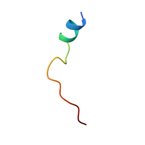
PROTEIN KINASE INHIBITOR PEPTIDEB [auth I]20Homo sapiensMutation(s): 0 
Gene Names: PKIA
UniProt & NIH Common Fund Data Resources
Find proteins for P61925 (Homo sapiens)
Explore P61925 
Go to UniProtKB:  P61925
PHAROS:  P61925
GTEx:  ENSG00000171033 
Entity Groups  
Sequence Clusters30% Identity50% Identity70% Identity90% Identity95% Identity100% Identity
UniProt GroupP61925
Sequence Annotations
Expand
  • Reference Sequence
Small Molecules
Ligands 1 Unique
IDChains Name / Formula / InChI Key2D Diagram3D Interactions
IQP
Query on IQP

Download Ideal Coordinates CCD File 
C [auth E]1-(5-ISOQUINOLINESULFONYL)-2-METHYLPIPERAZINE
C14 H17 N3 O2 S
BDVFVCGFMNCYPV-NSHDSACASA-N
Modified Residues  2 Unique
IDChains TypeFormula2D DiagramParent
SEP
Query on SEP
A [auth E]L-PEPTIDE LINKINGC3 H8 N O6 PSER
TPO
Query on TPO
A [auth E]L-PEPTIDE LINKINGC4 H10 N O6 PTHR
Binding Affinity Annotations 
IDSourceBinding Affinity
IQP PDBBind:  1YDR Ki: 3000 (nM) from 1 assay(s)
BindingDB:  1YDR Ki: 3000 (nM) from 1 assay(s)
IC50: 3000 (nM) from 1 assay(s)
Experimental Data & Validation

Experimental Data

  • Method: X-RAY DIFFRACTION
  • Resolution: 2.20 Å
  • R-Value Work: 0.188 
  • R-Value Observed: 0.188 
  • Space Group: P 21 21 21
Unit Cell:
Length ( Å )Angle ( ˚ )
a = 73.58α = 90
b = 76.28β = 90
c = 80.58γ = 90
Software Package:
Software NamePurpose
X-PLORmodel building
X-PLORrefinement
SAINTdata reduction
SAINTdata scaling
X-PLORphasing

Structure Validation

View Full Validation Report


Ligand Structure Quality Assessment 


Entry History 

Deposition Data

Revision History  (Full details and data files)

  • Version 1.0: 1997-04-01
    Type: Initial release
  • Version 1.1: 2008-03-24
    Changes: Version format compliance
  • Version 1.2: 2011-07-13
    Changes: Version format compliance
  • Version 1.3: 2024-06-05
    Changes: Data collection, Database references, Derived calculations, Other
  • Version 1.4: 2024-10-23
    Changes: Structure summary