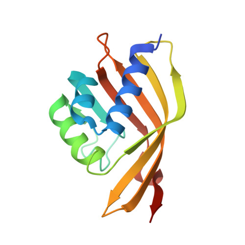
2A15

X-ray Crystal Structure of RV0760 from Mycobacterium Tuberculosis at 1.68 Angstrom Resolution


Experimental Data Snapshot

  • Method: X-RAY DIFFRACTION
  • Resolution: 1.68 Å
  • R-Value Free: 0.207 
  • R-Value Work: 0.176 
  • R-Value Observed: 0.178 

Starting Model: experimental
View more details

wwPDB Validation   3D Report Full Report


This is version 1.6 of the entry. See complete history


Literature

Crystal structure of Mycobacterium tuberculosis Rv0760c at 1.50 A resolution, a structural homolog of Delta(5)-3-ketosteroid isomerase

Cherney, M.M.Garen, C.R.James, M.N.G.

(2008) Biochim Biophys Acta 1784: 1625-1632

  • DOI: https://doi.org/10.1016/j.bbapap.2008.05.012
  • Primary Citation of Related Structures:  
    2A15, 2Z76, 2Z77, 2Z7A

  • PubMed Abstract: 

    We have determined the X-ray crystal structure of the Mycobacterium tuberculosis (Mtb) gene product encoded by the open reading frame Rv0760c at 1.50 A resolution by single-wavelength anomalous dispersion (SAD) phasing of diffraction data from crystals of the selenomethionine-substituted protein. Refinement against diffraction data from the native protein resulted in R(work)=19.5% and R(free)=21.4%. The X-ray crystal structure shows that the homodimeric Rv0760c polypeptide has an alpha + beta conical barrel fold placing it among many structural neighbors of the nuclear transport factor 2 family (NTF2). This family is highly conserved in terms of structure; however the substrates and individual protein functions are diverse. The structures of native Rv0760c in several different crystal forms and Rv0760c bound to 17beta-estradiol 17-hemisuccinate (EH) have also been solved and analyzed.


  • Organizational Affiliation

    Group in Protein Structure and Function, Department of Biochemistry, University of Alberta, Edmonton, Alberta, Canada T6G 2H7.


Macromolecules
Find similar proteins by:  (by identity cutoff)  |  3D Structure
Entity ID: 1
MoleculeChains Sequence LengthOrganismDetailsImage
HYPOTHETICAL PROTEIN Rv0760c139Mycobacterium tuberculosisMutation(s): 0 
Gene Names: Rv0760
UniProt
Find proteins for P71817 (Mycobacterium tuberculosis (strain CDC 1551 / Oshkosh))
Explore P71817 
Go to UniProtKB:  P71817
Entity Groups  
Sequence Clusters30% Identity50% Identity70% Identity90% Identity95% Identity100% Identity
UniProt GroupP71817
Sequence Annotations
Expand
  • Reference Sequence
Small Molecules
Ligands 1 Unique
IDChains Name / Formula / InChI Key2D Diagram3D Interactions
NCA
Query on NCA

Download Ideal Coordinates CCD File 
B [auth A]NICOTINAMIDE
C6 H6 N2 O
DFPAKSUCGFBDDF-UHFFFAOYSA-N
Experimental Data & Validation

Experimental Data

  • Method: X-RAY DIFFRACTION
  • Resolution: 1.68 Å
  • R-Value Free: 0.207 
  • R-Value Work: 0.176 
  • R-Value Observed: 0.178 
  • Space Group: P 32 1 2
Unit Cell:
Length ( Å )Angle ( ˚ )
a = 55.6α = 90
b = 55.6β = 90
c = 104.74γ = 120
Software Package:
Software NamePurpose
REFMACrefinement
SCALEPACKdata scaling
MOLREPphasing
ELVESrefinement
ELVESdata reduction

Structure Validation

View Full Validation Report


Entry History 

Deposition Data

Revision History  (Full details and data files)

  • Version 1.0: 2005-10-11
    Type: Initial release
  • Version 1.1: 2008-04-30
    Changes: Version format compliance
  • Version 1.2: 2011-07-13
    Changes: Version format compliance
  • Version 1.3: 2017-10-11
    Changes: Refinement description
  • Version 1.4: 2019-07-24
    Changes: Data collection, Refinement description
  • Version 1.5: 2024-02-14
    Changes: Data collection, Database references, Derived calculations
  • Version 1.6: 2024-04-03
    Changes: Refinement description