2FUE | pdb_00002fue

Human alpha-Phosphomannomutase 1 with D-mannose 1-phosphate and Mg2+ cofactor bound


Experimental Data Snapshot

  • Method: X-RAY DIFFRACTION
  • Resolution: 1.75 Å
  • R-Value Free: 
    0.240 (Depositor), 0.250 (DCC) 
  • R-Value Work: 
    0.202 (Depositor), 0.210 (DCC) 
  • R-Value Observed: 
    0.204 (Depositor) 

Starting Model: experimental
View more details

wwPDB Validation   3D Report Full Report


Ligand Structure Quality Assessment 

Created with Raphaël 2.3.0Worse 01 BetterLigand structure goodness of fit to experimental dataBest fitted M1PClick on this verticalbar to view details

This is version 1.7 of the entry. See complete history


Literature

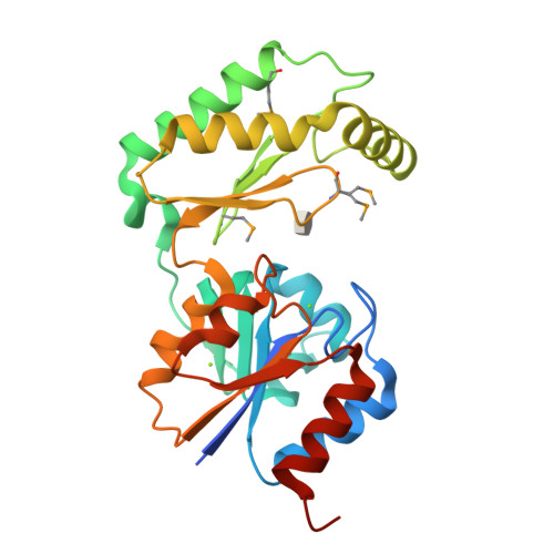
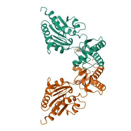
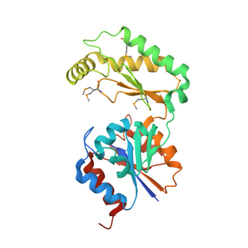
The X-ray crystal structures of human alpha-phosphomannomutase 1 reveal the structural basis of congenital disorder of glycosylation type 1a.

Silvaggi, N.R.Zhang, C.Lu, Z.Dai, J.Dunaway-Mariano, D.Allen, K.N.

(2006) J Biological Chem 281: 14918-14926

  • DOI: https://doi.org/10.1074/jbc.M601505200
  • Primary Citation of Related Structures:  
    2FUC, 2FUE

  • PubMed Abstract: 

    Congenital disorder of glycosylation type 1a (CDG-1a) is a congenital disease characterized by severe defects in nervous system development. It is caused by mutations in alpha-phosphomannomutase (of which there are two isozymes, alpha-PMM1 and alpha-PPM2). Here we report the x-ray crystal structures of human alpha-PMM1 in the open conformation, with and without the bound substrate, alpha-D-mannose 1-phosphate. Alpha-PMM1, like most haloalkanoic acid dehalogenase superfamily (HADSF) members, consists of two domains, the cap and core, which open to bind substrate and then close to provide a solvent-exclusive environment for catalysis. The substrate phosphate group is observed at a positively charged site of the cap domain, rather than at the core domain phosphoryl-transfer site defined by the Asp(19) nucleophile and Mg(2+) cofactor. This suggests that substrate binds first to the cap and then is swept into the active site upon cap closure. The orientation of the acid/base residue Asp(21) suggests that alpha-phosphomannomutase (alpha-PMM) uses a different method of protecting the aspartylphosphate from hydrolysis than the HADSF member beta-phosphoglucomutase. It is hypothesized that the electrostatic repulsion of positive charges at the interface of the cap and core domains stabilizes alpha-PMM1 in the open conformation and that the negatively charged substrate binds to the cap, thereby facilitating its closure over the core domain. The two isozymes, alpha-PMM1 and alpha-PMM2, are shown to have a conserved active-site structure and to display similar kinetic properties. Analysis of the known mutation sites in the context of the structures reveals the genotype-phenotype relationship underlying CDG-1a.


  • Organizational Affiliation

    Department of Physiology and Biophysics, Boston University School of Medicine, 715 Albany Street, Boston, MA 02118, USA.


Macromolecules
Find similar proteins by:  (by identity cutoff)  |  3D Structure
Entity ID: 1
MoleculeChains Sequence LengthOrganismDetailsImage
Phosphomannomutase 1262Homo sapiensMutation(s): 4 
EC: 5.4.2.8
UniProt & NIH Common Fund Data Resources
Find proteins for Q92871 (Homo sapiens)
Explore Q92871 
Go to UniProtKB:  Q92871
PHAROS:  Q92871
GTEx:  ENSG00000100417 
Entity Groups  
Sequence Clusters30% Identity50% Identity70% Identity90% Identity95% Identity100% Identity
UniProt GroupQ92871
Sequence Annotations
Expand
  • Reference Sequence
Experimental Data & Validation

Experimental Data

  • Method: X-RAY DIFFRACTION
  • Resolution: 1.75 Å
  • R-Value Free:  0.240 (Depositor), 0.250 (DCC) 
  • R-Value Work:  0.202 (Depositor), 0.210 (DCC) 
  • R-Value Observed: 0.204 (Depositor) 
Space Group: P 43 21 2
Unit Cell:
Length ( Å )Angle ( ˚ )
a = 51.39α = 90
b = 51.39β = 90
c = 214.603γ = 90
Software Package:
Software NamePurpose
DENZOdata reduction
SCALEPACKdata scaling
REFMACrefinement
PDB_EXTRACTdata extraction
Cootmodel building

Structure Validation

View Full Validation Report


Ligand Structure Quality Assessment 

Created with Raphaël 2.3.0Worse 01 BetterLigand structure goodness of fit to experimental dataBest fitted M1PClick on this verticalbar to view details

Entry History 

Deposition Data

Revision History  (Full details and data files)

  • Version 1.0: 2006-03-21
    Type: Initial release
  • Version 1.1: 2008-05-01
    Changes: Version format compliance
  • Version 1.2: 2011-07-13
    Changes: Advisory, Version format compliance
  • Version 1.3: 2017-10-18
    Changes: Advisory, Refinement description
  • Version 1.4: 2020-07-29
    Type: Remediation
    Reason: Carbohydrate remediation
    Changes: Data collection, Database references, Derived calculations, Structure summary
  • Version 1.5: 2023-08-30
    Changes: Advisory, Data collection, Database references, Refinement description, Structure summary
  • Version 1.6: 2023-11-15
    Changes: Data collection
  • Version 1.7: 2024-10-30
    Changes: Structure summary