2I54

Phosphomannomutase from Leishmania mexicana


Experimental Data Snapshot

  • Method: X-RAY DIFFRACTION
  • Resolution: 2.10 Å
  • R-Value Free: 0.230 
  • R-Value Work: 0.189 

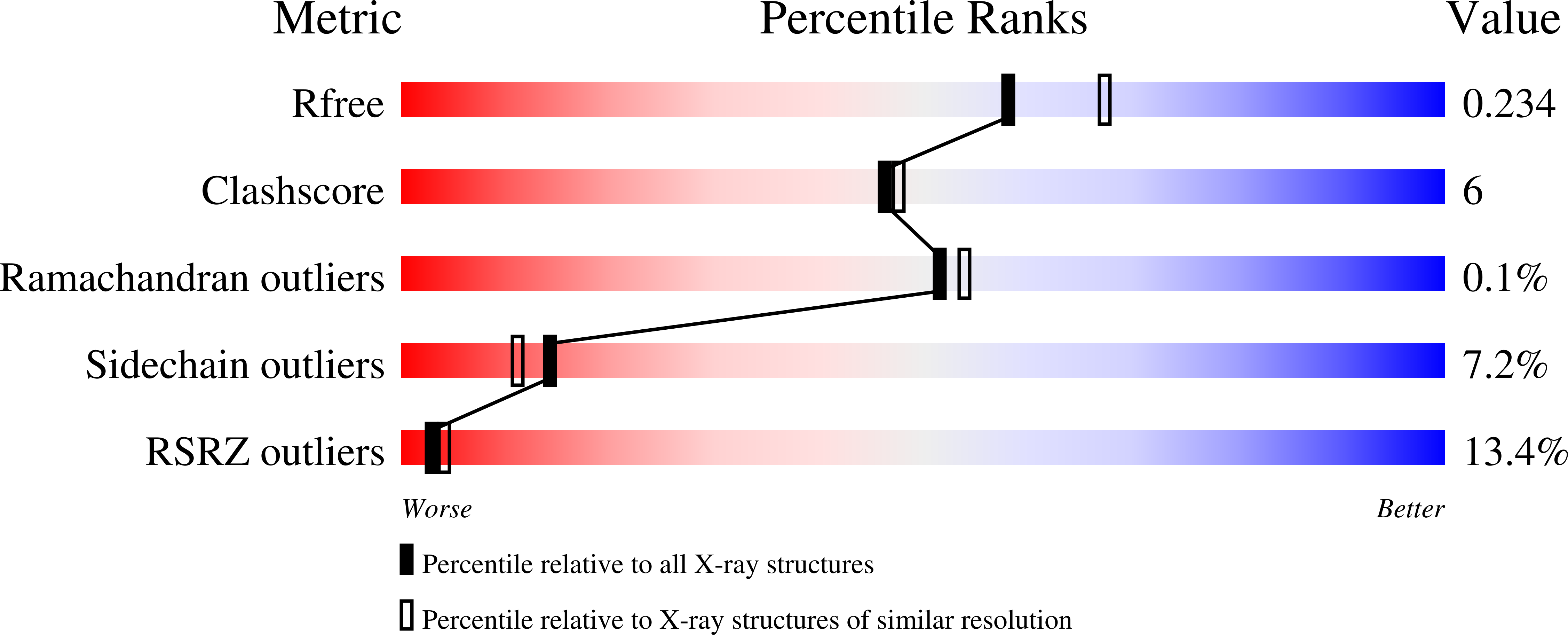
wwPDB Validation   3D Report Full Report


This is version 1.3 of the entry. See complete history


Literature

Structure of Leishmania mexicana phosphomannomutase highlights similarities with human isoforms

Kedzierski, L.Malby, R.L.Smith, B.J.Perugini, M.A.Hodder, A.N.Ilg, T.Colman, P.M.Handman, E.

(2006) J Mol Biol 363: 215-227

  • DOI: https://doi.org/10.1016/j.jmb.2006.08.023
  • Primary Citation of Related Structures:  
    2I54, 2I55

  • PubMed Abstract: 

    Phosphomannomutase (PMM) catalyses the conversion of mannose-6-phosphate to mannose-1-phosphate, an essential step in mannose activation and the biosynthesis of glycoconjugates in all eukaryotes. Deletion of PMM from Leishmania mexicana results in loss of virulence, suggesting that PMM is a promising drug target for the development of anti-leishmanial inhibitors. We report the crystallization and structure determination to 2.1 A of L. mexicana PMM alone and in complex with glucose-1,6-bisphosphate to 2.9 A. PMM is a member of the haloacid dehalogenase (HAD) family, but has a novel dimeric structure and a distinct cap domain of unique topology. Although the structure is novel within the HAD family, the leishmanial enzyme shows a high degree of similarity with its human isoforms. We have generated L. major PMM knockouts, which are avirulent. We expressed the human pmm2 gene in the Leishmania PMM knockout, but despite the similarity between Leishmania and human PMM, expression of the human gene did not restore virulence. Similarities in the structure of the parasite enzyme and its human isoforms suggest that the development of parasite-selective inhibitors will not be an easy task.


  • Organizational Affiliation

    Infection and Immunity Division, The Walter and Eliza Hall Institute of Medical Research, Melbourne, Australia.


Macromolecules
Find similar proteins by:  (by identity cutoff)  |  3D Structure
Entity ID: 1
MoleculeChains Sequence LengthOrganismDetailsImage
Phosphomannomutase
A, B, C
247Leishmania mexicanaMutation(s): 0 
EC: 5.4.2.8
UniProt
Find proteins for Q95ZD7 (Leishmania mexicana)
Explore Q95ZD7 
Go to UniProtKB:  Q95ZD7
Entity Groups  
Sequence Clusters30% Identity50% Identity70% Identity90% Identity95% Identity100% Identity
UniProt GroupQ95ZD7
Sequence Annotations
Expand
  • Reference Sequence
Small Molecules
Ligands 3 Unique
IDChains Name / Formula / InChI Key2D Diagram3D Interactions
CIT
Query on CIT

Download Ideal Coordinates CCD File 
F [auth A],
I [auth B],
K [auth C]
CITRIC ACID
C6 H8 O7
KRKNYBCHXYNGOX-UHFFFAOYSA-N
CL
Query on CL

Download Ideal Coordinates CCD File 
D [auth A],
G [auth B]
CHLORIDE ION
Cl
VEXZGXHMUGYJMC-UHFFFAOYSA-M
MG
Query on MG

Download Ideal Coordinates CCD File 
E [auth A],
H [auth B],
J [auth C]
MAGNESIUM ION
Mg
JLVVSXFLKOJNIY-UHFFFAOYSA-N
Experimental Data & Validation

Experimental Data

  • Method: X-RAY DIFFRACTION
  • Resolution: 2.10 Å
  • R-Value Free: 0.230 
  • R-Value Work: 0.189 
  • Space Group: P 31 2 1
Unit Cell:
Length ( Å )Angle ( ˚ )
a = 92.413α = 90
b = 92.413β = 90
c = 173.249γ = 120
Software Package:
Software NamePurpose
REFMACrefinement
PDB_EXTRACTdata extraction
HKL-2000data collection
HKL-2000data reduction
HKL-2000data scaling
SHARPphasing

Structure Validation

View Full Validation Report


Entry History 

Deposition Data

  • Released Date: 2007-04-17 
  • Deposition Author(s): Smith, B.J.

Revision History  (Full details and data files)

  • Version 1.0: 2007-04-17
    Type: Initial release
  • Version 1.1: 2008-05-01
    Changes: Version format compliance
  • Version 1.2: 2011-07-13
    Changes: Advisory, Version format compliance
  • Version 1.3: 2024-03-13
    Changes: Data collection, Database references, Derived calculations