2IGA | pdb_00002iga

Structure of Homoprotocatechuate 2,3-Dioxygenase from B. fuscum in complex with reactive intermediates formed via in crystallo reaction with 4-nitrocatechol at low oxygen concentrations.


Experimental Data Snapshot

  • Method: X-RAY DIFFRACTION
  • Resolution: 1.95 Å
  • R-Value Free: 
    0.245 (Depositor), 0.250 (DCC) 
  • R-Value Work: 
    0.185 (Depositor), 0.190 (DCC) 

Starting Model: experimental
View more details

wwPDB Validation   3D Report Full Report


Ligand Structure Quality Assessment 

Created with Raphaël 2.3.0Worse 01 BetterLigand structure goodness of fit to experimental dataBest fitted XX3Click on this verticalbar to view detailsBest fitted XXPClick on this verticalbar to view detailsBest fitted XX2Click on this verticalbar to view details

This is version 1.3 of the entry. See complete history


Literature

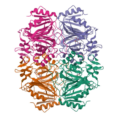
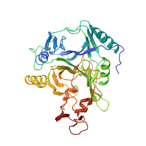
Crystal structures of Fe2+ dioxygenase superoxo, alkylperoxo, and bound product intermediates

Kovaleva, E.G.Lipscomb, J.D.

(2007) Science 316: 453-457

  • DOI: https://doi.org/10.1126/science.1134697
  • Primary Citation of Related Structures:  
    2IG9, 2IGA

  • PubMed Abstract: 

    We report the structures of three intermediates in the O2 activation and insertion reactions of an extradiol ring-cleaving dioxygenase. A crystal of Fe2+-containing homoprotocatechuate 2,3-dioxygenase was soaked in the slow substrate 4-nitrocatechol in a low O2 atmosphere. The x-ray crystal structure shows that three different intermediates reside in different subunits of a single homotetrameric enzyme molecule. One of these is the key substrate-alkylperoxo-Fe2+ intermediate, which has been predicted, but not structurally characterized, in an oxygenase. The intermediates define the major chemical steps of the dioxygenase mechanism and point to a general mechanistic strategy for the diverse 2-His-1-carboxylate enzyme family.


  • Organizational Affiliation

    Department of Biochemistry, Molecular Biology, and Biophysics, University of Minnesota, Minneapolis, MN 55455, USA.


Macromolecules
Find similar proteins by:  (by identity cutoff)  |  3D Structure
Entity ID: 1
MoleculeChains Sequence LengthOrganismDetailsImage
Homoprotocatechuate 2,3-dioxygenase
A, B, C, D
365Brevibacterium fuscumMutation(s): 0 
Gene Names: hpcd
EC: 1.13.11.15
UniProt
Find proteins for Q45135 (Brevibacterium fuscum)
Explore Q45135 
Go to UniProtKB:  Q45135
Entity Groups  
Sequence Clusters30% Identity50% Identity70% Identity90% Identity95% Identity100% Identity
UniProt GroupQ45135
Sequence Annotations
Expand
  • Reference Sequence
Small Molecules
Ligands 8 Unique
IDChains Name / Formula / InChI Key2D Diagram3D Interactions
XX3
Query on XX3

Download Ideal Coordinates CCD File 
CA [auth D],
M [auth B]
(1S)-1-HYDROPEROXY-1-HYDROXY-2-KETO-5-NITROCYCLOHEXA-3,5-DIENE
C6 H5 N O6
FWUJUIQFDWUIAR-LURJTMIESA-N
XXP
Query on XXP

Download Ideal Coordinates CCD File 
G [auth A]2-KETO,5-NITRO,6-HYDROXY-3,5-HEXADIENOIC ACID
C6 H5 N O6
UDOBAQIWFWMQMT-BXTBVDPRSA-N
XX2
Query on XX2

Download Ideal Coordinates CCD File 
V [auth C]1-KETO,2-HYDROXY,4-NITROBENZENE, 1 ELECTRON OXIDIZED
C6 H4 N O4
KJBVYJMPBKCQJZ-ZCFIWIBFSA-N
GOL
Query on GOL

Download Ideal Coordinates CCD File 
DA [auth D]
EA [auth D]
H [auth A]
I [auth A]
N [auth B]
GLYCEROL
C3 H8 O3
PEDCQBHIVMGVHV-UHFFFAOYSA-N
FE2
Query on FE2

Download Ideal Coordinates CCD File 
AA [auth D],
E [auth A],
J [auth B],
T [auth C]
FE (II) ION
Fe
CWYNVVGOOAEACU-UHFFFAOYSA-N
CA
Query on CA

Download Ideal Coordinates CCD File 
L [auth B]CALCIUM ION
Ca
BHPQYMZQTOCNFJ-UHFFFAOYSA-N
CL
Query on CL

Download Ideal Coordinates CCD File 
BA [auth D],
F [auth A],
K [auth B],
U [auth C]
CHLORIDE ION
Cl
VEXZGXHMUGYJMC-UHFFFAOYSA-M
OXY
Query on OXY

Download Ideal Coordinates CCD File 
W [auth C]OXYGEN MOLECULE
O2
MYMOFIZGZYHOMD-UHFFFAOYSA-N
Experimental Data & Validation

Experimental Data

  • Method: X-RAY DIFFRACTION
  • Resolution: 1.95 Å
  • R-Value Free:  0.245 (Depositor), 0.250 (DCC) 
  • R-Value Work:  0.185 (Depositor), 0.190 (DCC) 
Space Group: P 21 21 2
Unit Cell:
Length ( Å )Angle ( ˚ )
a = 110.678α = 90
b = 153.022β = 90
c = 96.389γ = 90
Software Package:
Software NamePurpose
REFMACrefinement
d*TREKdata reduction
d*TREKdata scaling
CCP4phasing

Structure Validation

View Full Validation Report


Ligand Structure Quality Assessment 

Created with Raphaël 2.3.0Worse 01 BetterLigand structure goodness of fit to experimental dataBest fitted XX3Click on this verticalbar to view detailsBest fitted XXPClick on this verticalbar to view detailsBest fitted XX2Click on this verticalbar to view details

Entry History 

Deposition Data

Revision History  (Full details and data files)

  • Version 1.0: 2007-04-24
    Type: Initial release
  • Version 1.1: 2007-10-16
    Changes: Version format compliance
  • Version 1.2: 2011-07-13
    Changes: Derived calculations, Version format compliance
  • Version 1.3: 2023-08-30
    Changes: Data collection, Database references, Derived calculations, Refinement description