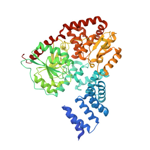
2JLB

Xanthomonas campestris putative OGT (XCC0866), complex with UDP- GlcNAc phosphonate analogue


Experimental Data Snapshot

  • Method: X-RAY DIFFRACTION
  • Resolution: 2.50 Å
  • R-Value Free: 0.247 
  • R-Value Work: 0.210 
  • R-Value Observed: 0.211 

Starting Model: experimental
View more details

wwPDB Validation   3D Report Full Report


Ligand Structure Quality Assessment 


This is version 1.2 of the entry. See complete history


Literature

Structural Insights Into Mechanism and Specificity of O-Glcnac Transferase.

Clarke, A.J.Hurtado-Guerrero, R.Pathak, S.Schuttelkopf, A.W.Borodkin, V.Shepherd, S.M.Ibrahim, A.F.M.Van Aalten, D.M.F.

(2008) EMBO J 27: 2780

  • DOI: https://doi.org/10.1038/emboj.2008.186
  • Primary Citation of Related Structures:  
    2JLB, 2VSY

  • PubMed Abstract: 

    Post-translational modification of protein serines/threonines with N-acetylglucosamine (O-GlcNAc) is dynamic, inducible and abundant, regulating many cellular processes by interfering with protein phosphorylation. O-GlcNAcylation is regulated by O-GlcNAc transferase (OGT) and O-GlcNAcase, both encoded by single, essential, genes in metazoan genomes. It is not understood how OGT recognises its sugar nucleotide donor and performs O-GlcNAc transfer onto proteins/peptides, and how the enzyme recognises specific cellular protein substrates. Here, we show, by X-ray crystallography and mutagenesis, that OGT adopts the (metal-independent) GT-B fold and binds a UDP-GlcNAc analogue at the bottom of a highly conserved putative peptide-binding groove, covered by a mobile loop. Strikingly, the tetratricopeptide repeats (TPRs) tightly interact with the active site to form a continuous 120 A putative interaction surface, whereas the previously predicted phosphatidylinositide-binding site locates to the opposite end of the catalytic domain. On the basis of the structure, we identify truncation/point mutants of the TPRs that have differential effects on activity towards proteins/peptides, giving first insights into how OGT may recognise its substrates.


  • Organizational Affiliation

    Division of Biological Chemistry & Drug Discovery, College of Life Sciences, University of Dundee, Dundee, UK.


Macromolecules
Find similar proteins by:  (by identity cutoff)  |  3D Structure
Entity ID: 1
MoleculeChains Sequence LengthOrganismDetailsImage
XCC0866
A, B
568Xanthomonas campestris pv. campestrisMutation(s): 0 
EC: 2.4.1.255
UniProt
Find proteins for Q8PC69 (Xanthomonas campestris pv. campestris (strain ATCC 33913 / DSM 3586 / NCPPB 528 / LMG 568 / P 25))
Explore Q8PC69 
Go to UniProtKB:  Q8PC69
Entity Groups  
Sequence Clusters30% Identity50% Identity70% Identity90% Identity95% Identity100% Identity
UniProt GroupQ8PC69
Sequence Annotations
Expand
  • Reference Sequence
Experimental Data & Validation

Experimental Data

  • Method: X-RAY DIFFRACTION
  • Resolution: 2.50 Å
  • R-Value Free: 0.247 
  • R-Value Work: 0.210 
  • R-Value Observed: 0.211 
  • Space Group: P 21 21 21
Unit Cell:
Length ( Å )Angle ( ˚ )
a = 83.86α = 90
b = 100.527β = 90
c = 154.521γ = 90
Software Package:
Software NamePurpose
REFMACrefinement
DENZOdata reduction
SCALEPACKdata scaling

Structure Validation

View Full Validation Report


Ligand Structure Quality Assessment 


Entry History 

Deposition Data

Revision History  (Full details and data files)

  • Version 1.0: 2008-11-25
    Type: Initial release
  • Version 1.1: 2011-07-13
    Changes: Advisory, Version format compliance
  • Version 1.2: 2023-12-13
    Changes: Data collection, Database references, Derived calculations, Other, Refinement description