2UVW

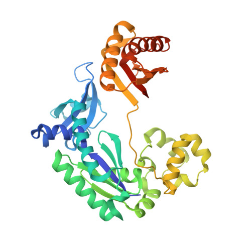
Crystal structures of mutant Dpo4 DNA polymerases with 8-oxoG containing DNA template-primer constructs


Experimental Data Snapshot

  • Method: X-RAY DIFFRACTION
  • Resolution: 2.09 Å
  • R-Value Free: 0.274 
  • R-Value Work: 0.249 
  • R-Value Observed: 0.249 

Starting Model: experimental
View more details

wwPDB Validation   3D Report Full Report


Ligand Structure Quality Assessment 


This is version 1.3 of the entry. See complete history


Literature

Hydrogen Bonding of 7,8-Dihydro-8-Oxodeoxyguanosine with a Charged Residue in the Little Finger Domain Determines Miscoding Events in Sulfolobus Solfataricus DNA Polymerase Dpo4.

Eoff, R.L.Irimia, A.Angel, K.C.Egli, M.Guengerich, F.P.

(2007) J Biol Chem 282: 19831

  • DOI: https://doi.org/10.1074/jbc.M702290200
  • Primary Citation of Related Structures:  
    2UVR, 2UVU, 2UVV, 2UVW

  • PubMed Abstract: 

    Sulfolobus solfataricus P2 DNA polymerase IV (Dpo4) has been shown to catalyze bypass of 7,8-dihydro-8-oxodeoxyguanosine (8-oxoG) in a highly efficient and relatively accurate manner. Crystal structures have revealed a potential role for Arg(332) in stabilizing the anti conformation of the 8-oxoG template base by means of a hydrogen bond or ion-dipole pair, which results in an increased enzymatic efficiency for dCTP insertion and makes formation of a Hoogsteen pair between 8-oxoG and dATP less favorable. Site-directed mutagenesis was used to replace Arg(332) with Ala, Glu, Leu, or His in order to probe the importance of Arg(332) in accurate and efficient bypass of 8-oxoG. The double mutant Ala(331)Ala(332) was also prepared to address the contribution of Arg(331). Transientstate kinetic results suggest that Glu(332) retains fidelity against bypass of 8-oxoG that is similar to wild type Dpo4, a result that was confirmed by tandem mass spectrometric analysis of full-length extension products. A crystal structure of the Dpo4 Glu(332) mutant and 8-oxoG:C pair revealed water-mediated hydrogen bonds between Glu(332) and the O-8 atom of 8-oxoG. The space normally occupied by Arg(332) side chain is empty in the crystal structures of the Ala(332) mutant. Two other crystal structures show that a Hoogsteen base pair is formed between 8-oxoG and A in the active site of both Glu(332) and Ala(332) mutants. These results support the view that a bond between Arg(332) and 8-oxoG plays a role in determining the fidelity and efficiency of Dpo4-catalyzed bypass of the lesion.


  • Organizational Affiliation

    Department of Biochemistry and Center in Molecular Toxicology, Vanderbilt University School of Medicine, Nashville, Tennessee 37232-0146, USA.


Macromolecules

Find similar proteins by:  (by identity cutoff)  |  3D Structure
Entity ID: 1
MoleculeChains Sequence LengthOrganismDetailsImage
DNA POLYMERASE IV358Saccharolobus solfataricus P2Mutation(s): 1 
EC: 2.7.7.7
UniProt
Find proteins for Q97W02 (Saccharolobus solfataricus (strain ATCC 35092 / DSM 1617 / JCM 11322 / P2))
Explore Q97W02 
Go to UniProtKB:  Q97W02
Entity Groups  
Sequence Clusters30% Identity50% Identity70% Identity90% Identity95% Identity100% Identity
UniProt GroupQ97W02
Sequence Annotations
Expand
  • Reference Sequence

Find similar nucleic acids by:  Sequence   |   3D Structure  

Entity ID: 2
MoleculeChains LengthOrganismImage
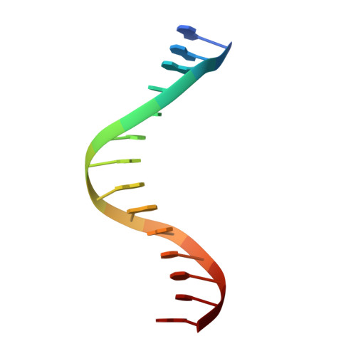
5'-D(*GP*GP*GP*GP*GP*AP*AP*GP*GP*AP *TP*TP*CP*A)-3'B [auth P]14N/A
Sequence Annotations
Expand
  • Reference Sequence

Find similar nucleic acids by:  Sequence   |   3D Structure  

Entity ID: 3
MoleculeChains LengthOrganismImage
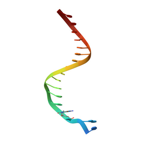
5'-D(*TP*CP*AP*CP*8OGP*GP*AP*AP*TP*CP *CP*TP*TP*CP*CP*CP*CP*C)-3'C [auth T]18N/A
Sequence Annotations
Expand
  • Reference Sequence
Experimental Data & Validation

Experimental Data

  • Method: X-RAY DIFFRACTION
  • Resolution: 2.09 Å
  • R-Value Free: 0.274 
  • R-Value Work: 0.249 
  • R-Value Observed: 0.249 
  • Space Group: P 21 21 2
Unit Cell:
Length ( Å )Angle ( ˚ )
a = 96.778α = 90
b = 103.728β = 90
c = 52.915γ = 90
Software Package:
Software NamePurpose
CNSrefinement
X-GENdata reduction
X-GENdata scaling
CNSphasing

Structure Validation

View Full Validation Report


Ligand Structure Quality Assessment 


Entry History 

Deposition Data

Revision History  (Full details and data files)

  • Version 1.0: 2007-05-08
    Type: Initial release
  • Version 1.1: 2011-05-08
    Changes: Version format compliance
  • Version 1.2: 2011-07-13
    Changes: Version format compliance
  • Version 1.3: 2023-12-13
    Changes: Data collection, Database references, Derived calculations, Other, Refinement description