2UWH

Cytochrome P450 BM3 mutant in complex with palmitic acid


Experimental Data Snapshot

  • Method: X-RAY DIFFRACTION
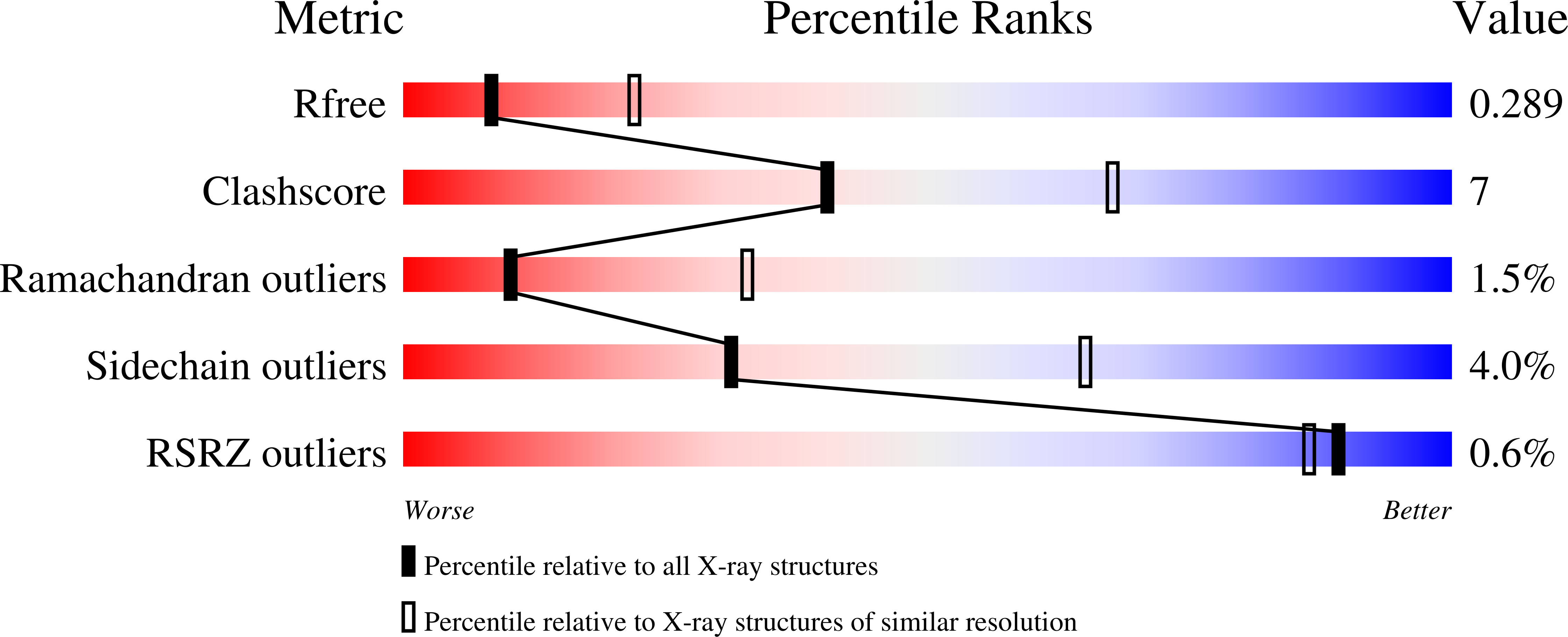
  • Resolution: 2.80 Å
  • R-Value Free: 0.299 
  • R-Value Work: 0.215 
  • R-Value Observed: 0.219 

Starting Model: experimental
View more details

wwPDB Validation   3D Report Full Report


Ligand Structure Quality Assessment 


This is version 1.3 of the entry. See complete history


Literature

Filling a Hole in Cytochrome P450 Bm3 Improves Substrate Binding and Catalytic Efficiency.

Huang, W.-C.Westlake, A.C.G.Marechal, J.-D.Joyce, M.G.Moody, P.C.E.Roberts, G.C.K.

(2007) J Mol Biol 373: 633

  • DOI: https://doi.org/10.1016/j.jmb.2007.08.015
  • Primary Citation of Related Structures:  
    2UWH

  • PubMed Abstract: 

    Cytochrome P450BM3 (CYP102A1) from Bacillus megaterium, a fatty acid hydroxylase, is a member of a very large superfamily of monooxygenase enzymes. The available crystal structures of the enzyme show non-productive binding of substrates with their omega-end distant from the iron in a hydrophobic pocket at one side of the active site. We have constructed and characterised mutants in which this pocket is filled by large hydrophobic side-chains replacing alanine at position 82. The mutants having phenylalanine or tryptophan at this position have very much (approximately 800-fold) greater affinity for substrate, with a greater conversion of the haem iron to the high-spin state, and similarly increased catalytic efficiency. The enzyme as isolated contains bound palmitate, reflecting this much higher affinity. We have determined the crystal structure of the haem domain of the Ala82Phe mutant with bound palmitate; this shows that the substrate is binding differently from the wild-type enzyme but still distant from the haem iron. Detailed analysis of the structure indicates that the tighter binding in the mutant reflects a shift in the conformational equilibrium of the substrate-free enzyme towards the conformation seen in the substrate complex rather than differences in the enzyme-substrate interactions. On this basis, we outline a sequence of events for the initial stages of the catalytic cycle. The Ala82Phe and Ala82Trp mutants are also very much more effective catalysts of indole hydroxylation than the wild-type enzyme, suggesting that they will be valuable starting points for the design of mutants to catalyse synthetically useful hydroxylation reactions.


  • Organizational Affiliation

    Henry Wellcome Laboratories of Structural Biology, Department of Biochemistry, University of Leicester, Leicester LE1 9HN, UK.


Macromolecules
Find similar proteins by:  (by identity cutoff)  |  3D Structure
Entity ID: 1
MoleculeChains Sequence LengthOrganismDetailsImage
BIFUNCTIONAL P-450\: NADPH-P450 REDUCTASE
A, B, C, D, E
A, B, C, D, E, F
458Priestia megateriumMutation(s): 1 
EC: 1.14.14.1 (PDB Primary Data), 1.6.2.4 (UniProt)
UniProt
Find proteins for P14779 (Priestia megaterium (strain ATCC 14581 / DSM 32 / CCUG 1817 / JCM 2506 / NBRC 15308 / NCIMB 9376 / NCTC 10342 / NRRL B-14308 / VKM B-512 / Ford 19))
Explore P14779 
Go to UniProtKB:  P14779
Entity Groups  
Sequence Clusters30% Identity50% Identity70% Identity90% Identity95% Identity100% Identity
UniProt GroupP14779
Sequence Annotations
Expand
  • Reference Sequence
Small Molecules
Ligands 2 Unique
IDChains Name / Formula / InChI Key2D Diagram3D Interactions
HEM
Query on HEM

Download Ideal Coordinates CCD File 
G [auth A]
I [auth B]
K [auth C]
M [auth D]
O [auth E]
G [auth A],
I [auth B],
K [auth C],
M [auth D],
O [auth E],
Q [auth F]
PROTOPORPHYRIN IX CONTAINING FE
C34 H32 Fe N4 O4
KABFMIBPWCXCRK-RGGAHWMASA-L
PLM
Query on PLM

Download Ideal Coordinates CCD File 
H [auth A]
J [auth B]
L [auth C]
N [auth D]
P [auth E]
H [auth A],
J [auth B],
L [auth C],
N [auth D],
P [auth E],
R [auth F]
PALMITIC ACID
C16 H32 O2
IPCSVZSSVZVIGE-UHFFFAOYSA-N
Experimental Data & Validation

Experimental Data

  • Method: X-RAY DIFFRACTION
  • Resolution: 2.80 Å
  • R-Value Free: 0.299 
  • R-Value Work: 0.215 
  • R-Value Observed: 0.219 
  • Space Group: P 21 21 21
Unit Cell:
Length ( Å )Angle ( ˚ )
a = 116.827α = 90
b = 147.03β = 90
c = 183.402γ = 90
Software Package:
Software NamePurpose
REFMACrefinement
MOSFLMdata reduction
SCALAdata scaling
PHASERphasing

Structure Validation

View Full Validation Report


Ligand Structure Quality Assessment 


Entry History 

Deposition Data

Revision History  (Full details and data files)

  • Version 1.0: 2007-08-28
    Type: Initial release
  • Version 1.1: 2011-05-08
    Changes: Version format compliance
  • Version 1.2: 2011-07-13
    Changes: Version format compliance
  • Version 1.3: 2023-12-13
    Changes: Data collection, Database references, Derived calculations, Other, Refinement description