Structural Basis for Complement Factor H Linked Age-Related Macular Degeneration.
Prosser, B.E., Johnson, S., Roversi, P., Herbert, A.P., Blaum, B.S., Tyrrell, J., Jowitt, T.A., Clark, S.J., Tarelli, E., Uhrin, D., Barlow, P.N., Sim, R.B., Day, A.J., Lea, S.M.(2007) J Exp Medicine 204: 2277
- PubMed: 17893204
- DOI: https://doi.org/10.1084/jem.20071069
- Primary Citation of Related Structures:
2UWN, 2V8E - PubMed Abstract:
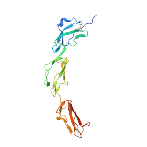
Nearly 50 million people worldwide suffer from age-related macular degeneration (AMD), which causes severe loss of central vision. A single-nucleotide polymorphism in the gene for the complement regulator factor H (FH), which causes a Tyr-to-His substitution at position 402, is linked to approximately 50% of attributable risks for AMD. We present the crystal structure of the region of FH containing the polymorphic amino acid His402 in complex with an analogue of the glycosaminoglycans (GAGs) that localize the complement regulator on the cell surface. The structure demonstrates direct coordination of ligand by the disease-associated polymorphic residue, providing a molecular explanation of the genetic observation. This glycan-binding site occupies the center of an extended interaction groove on the regulator's surface, implying multivalent binding of sulfated GAGs. This finding is confirmed by structure-based site-directed mutagenesis, nuclear magnetic resonance-monitored binding experiments performed for both H402 and Y402 variants with this and another model GAG, and analysis of an extended GAG-FH complex.
Organizational Affiliation:
Sir William Dunn School of Pathology, University of Oxford, Oxford, OX1 3RE, England, UK.


