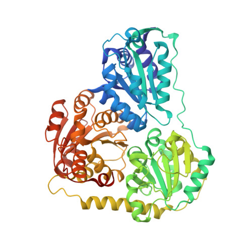
2UZ1

1.65 Angstrom structure of Benzaldehyde Lyase complexed with 2-methyl- 2,4-pentanediol


Experimental Data Snapshot

  • Method: X-RAY DIFFRACTION
  • Resolution: 1.65 Å
  • R-Value Free: 0.192 
  • R-Value Work: 0.167 
  • R-Value Observed: 0.168 

Starting Model: experimental
View more details

wwPDB Validation   3D Report Full Report


Ligand Structure Quality Assessment 


This is version 1.3 of the entry. See complete history


Literature

Structure of the Thdp-Dependent Enzyme Benzaldehyde Lyase Refined to 1.65 A Resolution.

Maraite, A.Schmidt, T.Ansorge-Schumacher, M.B.Brzozowski, A.M.Grogan, G.

(2007) Acta Crystallogr Sect F Struct Biol Cryst Commun 63: 546

  • DOI: https://doi.org/10.1107/S1744309107028576
  • Primary Citation of Related Structures:  
    2UZ1

  • PubMed Abstract: 

    Benzaldehyde lyase (BAL; EC 4.1.2.38) is a thiamine diphosphate (ThDP) dependent enzyme that catalyses the enantioselective carboligation of two molecules of benzaldehyde to form (R)-benzoin. BAL has hence aroused interest for its potential in the industrial synthesis of optically active benzoins and derivatives. The structure of BAL was previously solved to a resolution of 2.6 A using MAD experiments on a selenomethionine derivative [Mosbacher et al. (2005), FEBS J. 272, 6067-6076]. In this communication of parallel studies, BAL was crystallized in an alternative space group (P2(1)2(1)2(1)) and its structure refined to a resolution of 1.65 A, allowing detailed observation of the water structure, active-site interactions with ThDP and also the electron density for the co-solvent 2-methyl-2,4-pentanediol (MPD) at hydrophobic patches of the enzyme surface.


  • Organizational Affiliation

    Department of Biotechnology, Faculty of Natural Sciences, RWTH Aachen University, Worringerweg 1, 52074 Aachen, Germany.


Macromolecules
Find similar proteins by:  (by identity cutoff)  |  3D Structure
Entity ID: 1
MoleculeChains Sequence LengthOrganismDetailsImage
BENZALDEHYDE LYASE
A, D
563Pseudomonas fluorescensMutation(s): 0 
EC: 4.1.2.38
UniProt
Find proteins for Q9F4L3 (Pseudomonas fluorescens)
Explore Q9F4L3 
Go to UniProtKB:  Q9F4L3
Entity Groups  
Sequence Clusters30% Identity50% Identity70% Identity90% Identity95% Identity100% Identity
UniProt GroupQ9F4L3
Sequence Annotations
Expand
  • Reference Sequence
Find similar proteins by:  (by identity cutoff)  |  3D Structure
Entity ID: 2
MoleculeChains Sequence LengthOrganismDetailsImage
BENZALDEHYDE LYASE
B, C
563Pseudomonas fluorescensMutation(s): 0 
EC: 4.1.2.38
UniProt
Find proteins for Q9F4L3 (Pseudomonas fluorescens)
Explore Q9F4L3 
Go to UniProtKB:  Q9F4L3
Entity Groups  
Sequence Clusters30% Identity50% Identity70% Identity90% Identity95% Identity100% Identity
UniProt GroupQ9F4L3
Sequence Annotations
Expand
  • Reference Sequence
Experimental Data & Validation

Experimental Data

  • Method: X-RAY DIFFRACTION
  • Resolution: 1.65 Å
  • R-Value Free: 0.192 
  • R-Value Work: 0.167 
  • R-Value Observed: 0.168 
  • Space Group: P 21 21 21
Unit Cell:
Length ( Å )Angle ( ˚ )
a = 104.462α = 90
b = 121.601β = 90
c = 162.957γ = 90
Software Package:
Software NamePurpose
REFMACrefinement
DENZOdata reduction
SCALEPACKdata scaling
MOLREPphasing

Structure Validation

View Full Validation Report


Ligand Structure Quality Assessment 


Entry History 

Deposition Data

Revision History  (Full details and data files)

  • Version 1.0: 2007-07-24
    Type: Initial release
  • Version 1.1: 2011-05-08
    Changes: Version format compliance
  • Version 1.2: 2011-07-13
    Changes: Version format compliance
  • Version 1.3: 2023-12-13
    Changes: Data collection, Database references, Other, Refinement description