2VQ5 | pdb_00002vq5

X-ray structure of Norcoclaurine synthase from Thalictrum flavum in complex with dopamine and hydroxybenzaldehyde


Experimental Data Snapshot

  • Method: X-RAY DIFFRACTION
  • Resolution: 2.09 Å
  • R-Value Free: 
    0.266 (Depositor), 0.270 (DCC) 
  • R-Value Work: 
    0.221 (Depositor), 0.220 (DCC) 
  • R-Value Observed: 
    0.223 (Depositor) 

Starting Model: experimental
View more details

wwPDB Validation   3D Report Full Report


Ligand Structure Quality Assessment 

Created with Raphaël 2.3.0Worse 01 BetterLigand structure goodness of fit to experimental dataBest fitted LDPClick on this verticalbar to view details

This is version 1.4 of the entry. See complete history


Literature

Structural Basis of Enzymatic S-Norcoclaurine Biosynthesis.

Ilari, A.Franceschini, S.Bonamore, A.Arenghi, F.Botta, B.Macone, A.Pasquo, A.Bellucci, L.Boffi, A.

(2009) J Biological Chem 284: 897

  • DOI: https://doi.org/10.1074/jbc.M803738200
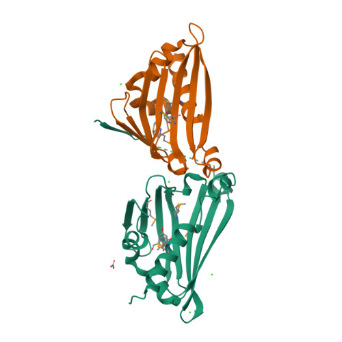
  • Primary Citation of Related Structures:  
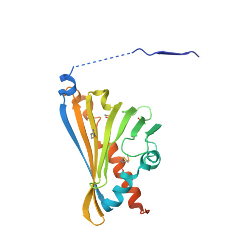
    2VNE, 2VQ5

  • PubMed Abstract: 

    The enzyme norcoclaurine synthase (NCS) catalyzes the stereospecific Pictet-Spengler cyclization between dopamine and 4-hydroxyphenylacetaldehyde, the key step in the benzylisoquinoline alkaloid biosynthetic pathway. The crystallographic structure of norcoclaurine synthase from Thalictrum flavum in its complex with dopamine substrate and the nonreactive substrate analogue 4-hydroxybenzaldehyde has been solved at 2.1A resolution. NCS shares no common features with the functionally correlated "Pictet-Spenglerases" that catalyze the first step of the indole alkaloids pathways and conforms to the overall fold of the Bet v1-like protein. The active site of NCS is located within a 20-A-long catalytic tunnel and is shaped by the side chains of a tyrosine, a lysine, an aspartic, and a glutamic acid. The geometry of the amino acid side chains with respect to the substrates reveals the structural determinants that govern the mechanism of the stereoselective Pictet-Spengler cyclization, thus establishing an excellent foundation for the understanding of the finer details of the catalytic process. Site-directed mutations of the relevant residues confirm the assignment based on crystallographic findings.


  • Organizational Affiliation

    Dipartimento di Scienze Biochimiche and Istituto di Biologia e Patologia Molecolari, CNR, Sapienza University, Piazzale Aldo Moro 5, 00185 Roma, Italy.


Macromolecules
Find similar proteins by:  (by identity cutoff)  |  3D Structure
Entity ID: 1
MoleculeChains Sequence LengthOrganismDetailsImage
S-NORCOCLAURINE SYNTHASE
A, B
201Thalictrum flavumMutation(s): 4 
EC: 4.2.1.78 (PDB Primary Data), 3.5.99.14 (UniProt)
Membrane Entity: Yes 
UniProt
Find proteins for Q67A25 (Thalictrum flavum subsp. glaucum)
Explore Q67A25 
Go to UniProtKB:  Q67A25
Entity Groups  
Sequence Clusters30% Identity50% Identity70% Identity90% Identity95% Identity100% Identity
UniProt GroupQ67A25
Sequence Annotations
Expand
  • Reference Sequence
Small Molecules
Ligands 4 Unique
IDChains Name / Formula / InChI Key2D Diagram3D Interactions
LDP
Query on LDP

Download Ideal Coordinates CCD File 
K [auth B]L-DOPAMINE
C8 H11 N O2
VYFYYTLLBUKUHU-UHFFFAOYSA-N
HBA
Query on HBA

Download Ideal Coordinates CCD File 
H [auth A],
L [auth B]
P-HYDROXYBENZALDEHYDE
C7 H6 O2
RGHHSNMVTDWUBI-UHFFFAOYSA-N
ACT
Query on ACT

Download Ideal Coordinates CCD File 
C [auth A],
D [auth A]
ACETATE ION
C2 H3 O2
QTBSBXVTEAMEQO-UHFFFAOYSA-M
CL
Query on CL

Download Ideal Coordinates CCD File 
E [auth A],
F [auth A],
G [auth A],
I [auth B],
J [auth B]
CHLORIDE ION
Cl
VEXZGXHMUGYJMC-UHFFFAOYSA-M
Modified Residues  1 Unique
IDChains TypeFormula2D DiagramParent
MSE
Query on MSE
A, B
L-PEPTIDE LINKINGC5 H11 N O2 SeMET
Experimental Data & Validation

Experimental Data

  • Method: X-RAY DIFFRACTION
  • Resolution: 2.09 Å
  • R-Value Free:  0.266 (Depositor), 0.270 (DCC) 
  • R-Value Work:  0.221 (Depositor), 0.220 (DCC) 
  • R-Value Observed: 0.223 (Depositor) 
Space Group: P 31 2 1
Unit Cell:
Length ( Å )Angle ( ˚ )
a = 86.474α = 90
b = 86.474β = 90
c = 117.977γ = 120
Software Package:
Software NamePurpose
REFMACrefinement
DENZOdata reduction
SCALEPACKdata scaling
MOLREPphasing

Structure Validation

View Full Validation Report


Ligand Structure Quality Assessment 

Created with Raphaël 2.3.0Worse 01 BetterLigand structure goodness of fit to experimental dataBest fitted LDPClick on this verticalbar to view details

Entry History 

Deposition Data

Revision History  (Full details and data files)

  • Version 1.0: 2008-08-05
    Type: Initial release
  • Version 1.1: 2011-05-08
    Changes: Version format compliance
  • Version 1.2: 2011-07-13
    Changes: Version format compliance
  • Version 1.3: 2023-12-13
    Changes: Data collection, Database references, Derived calculations, Other, Refinement description
  • Version 1.4: 2024-11-20
    Changes: Structure summary