Allosteric Activation of Pyruvate Decarboxylases. A Never-Ending Story.
Konig, S., Spinka, M., Kutter, S.(2014) J Mol Catal B Enzym 61: 100
Experimental Data Snapshot
Starting Model: experimental
View more details
(2014) J Mol Catal B Enzym 61: 100
Entity ID: 1 | |||||
|---|---|---|---|---|---|
| Molecule | Chains | Sequence Length | Organism | Details | Image |
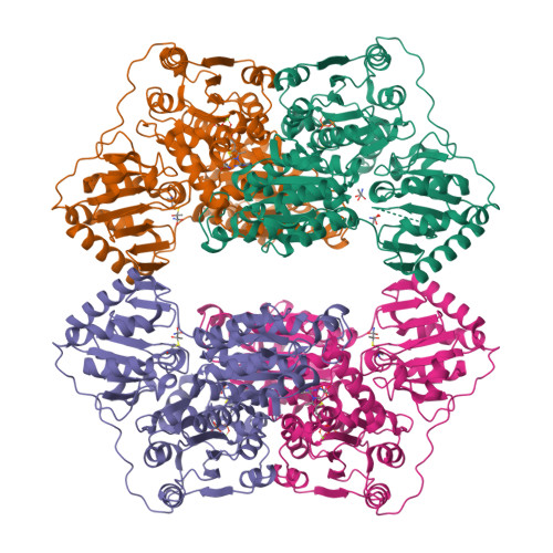
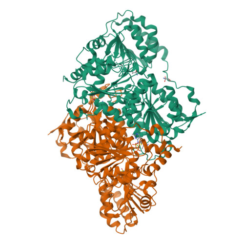
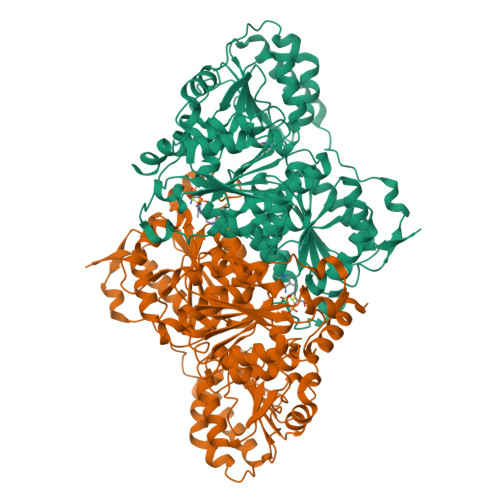
| PYRUVATE DECARBOXYLASE ISOZYME 1 | 563 | Saccharomyces cerevisiae | Mutation(s): 1 EC: 4.1.1.1 (PDB Primary Data), 4.1.1.74 (UniProt), 4.1.1.72 (UniProt), 4.1.1.43 (UniProt), 4.1.1 (UniProt) | 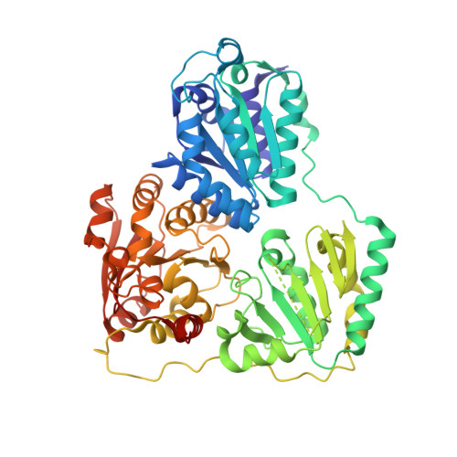 | |
UniProt | |||||
Find proteins for P06169 (Saccharomyces cerevisiae (strain ATCC 204508 / S288c)) Explore P06169 Go to UniProtKB: P06169 | |||||
Entity Groups | |||||
| Sequence Clusters | 30% Identity50% Identity70% Identity90% Identity95% Identity100% Identity | ||||
| UniProt Group | P06169 | ||||
Sequence AnnotationsExpand | |||||
| |||||
| Ligands 3 Unique | |||||
|---|---|---|---|---|---|
| ID | Chains | Name / Formula / InChI Key | 2D Diagram | 3D Interactions | |
| TPP Query on TPP | E [auth A], I [auth B], M [auth C], Q [auth D] | THIAMINE DIPHOSPHATE C12 H19 N4 O7 P2 S AYEKOFBPNLCAJY-UHFFFAOYSA-O |  | ||
| PY0 Query on PY0 | G [auth A] H [auth A] K [auth B] L [auth B] O [auth C] | (1S,2S)-1-amino-1,2-dihydroxypropan-1-olate C3 H8 N O3 ISJWJWXATCJUBY-STHAYSLISA-N |  | ||
| MG Query on MG | F [auth A], J [auth B], N [auth C], R [auth D] | MAGNESIUM ION Mg JLVVSXFLKOJNIY-UHFFFAOYSA-N |  | ||
| Length ( Å ) | Angle ( ˚ ) |
|---|---|
| a = 73.05 | α = 89.23 |
| b = 79.22 | β = 73.32 |
| c = 109.09 | γ = 62.43 |
| Software Name | Purpose |
|---|---|
| REFMAC | refinement |
| DENZO | data reduction |
| SCALEPACK | data scaling |
| MOLREP | phasing |