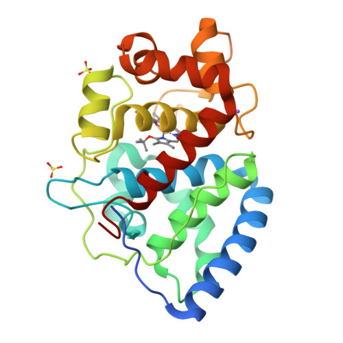
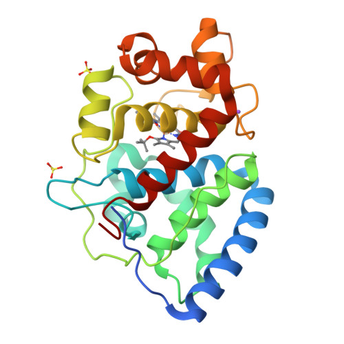
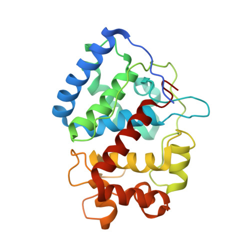
2WD4

Ascorbate Peroxidase as a heme oxygenase: w41A variant product with t-butyl peroxide


Experimental Data Snapshot

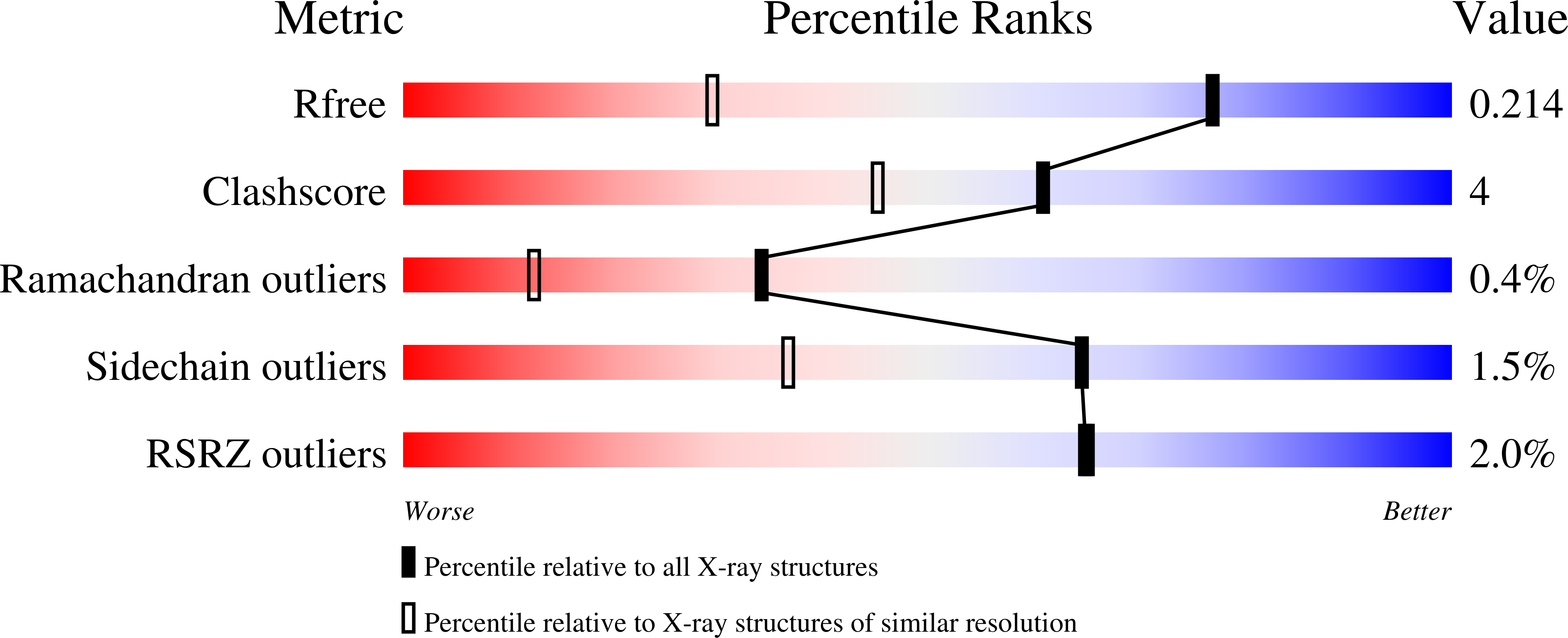
  • Method: X-RAY DIFFRACTION
  • Resolution: 1.40 Å
  • R-Value Free: 0.216 
  • R-Value Work: 0.190 
  • R-Value Observed: 0.191 

Starting Model: experimental
View more details

wwPDB Validation   3D Report Full Report


Ligand Structure Quality Assessment 


This is version 1.3 of the entry. See complete history


Literature

Evidence for Heme Oxygenase Activity in a Heme Peroxidase.

Raven, E.L.Badyal, S.K.Eaton, G.Mistry, S.Pipirou, Z.Basran, J.Metcalfe, C.L.Gumiero, A.Handa, S.Moody, P.C.E.

(2009) Biochemistry 48: 4738

  • DOI: https://doi.org/10.1021/bi900118j
  • Primary Citation of Related Structures:  
    2WD4

  • PubMed Abstract: 

    The heme peroxidase and heme oxygenase enzymes share a common heme prosthetic group but catalyze fundamentally different reactions, the first being H(2)O(2)-dependent oxidation of substrate using an oxidized Compound I intermediate, and the second O(2)-dependent degradation of heme. It has been proposed that these enzymes utilize a common reaction intermediate, a ferric hydroperoxide species, that sits at a crossroads in the mechanism and beyond which there are two mutually exclusive mechanistic pathways. Here, we present evidence to support this proposal in a heme peroxidase. Hence, we describe kinetic data for a variant of ascorbate peroxidase (W41A) which reacts slowly with tert-butyl hydroperoxide and does not form the usual peroxidase Compound I intermediate; instead, structural data show that a product is formed in which the heme has been cleaved at the alpha-meso position, analogous to the heme oxygenase mechanism. We interpret this to mean that the Compound I (peroxidase) pathway is shut down, so that instead the reaction intermediate diverts through the alternative (heme oxygenase) route. A mechanism for formation of the product is proposed and discussed in the light of what is known about the heme oxygenase reaction mechanism.


  • Organizational Affiliation

    Department of Chemistry, Henry Wellcome Building, University of Leicester, University Road, Leicester LE1 7RH, UK.


Macromolecules
Find similar proteins by:  (by identity cutoff)  |  3D Structure
Entity ID: 1
MoleculeChains Sequence LengthOrganismDetailsImage
ASCORBATE PEROXIDASE261Glycine maxMutation(s): 1 
EC: 1.11.1.11
UniProt
Find proteins for Q43758 (Glycine max)
Explore Q43758 
Go to UniProtKB:  Q43758
Entity Groups  
Sequence Clusters30% Identity50% Identity70% Identity90% Identity95% Identity100% Identity
UniProt GroupQ43758
Sequence Annotations
Expand
  • Reference Sequence
Small Molecules
Ligands 4 Unique
IDChains Name / Formula / InChI Key2D Diagram3D Interactions
TBV
Query on TBV

Download Ideal Coordinates CCD File 
E [auth A]3-[2-[[3-(2-CARBOXYETHYL)-5-[[3-ETHENYL-4-METHYL-5-[(2-METHYLPROPAN-2-YL)OXY]-1H-PYRROL-2-YL]METHYL]-4-METHYL-1H-PYRROL -2-YL]METHYL]-5-[(Z)-(4-ETHENYL-3-METHYL-5-OXO-PYRROL-2-YLIDENE)METHYL]-4-METHYL-1H-PYRROL-3-YL]PROPANOIC ACID
C37 H46 N4 O6
JDQNAGCTUKHXGS-MWLSYYOVSA-N
SO4
Query on SO4

Download Ideal Coordinates CCD File 
D [auth A],
F [auth A]
SULFATE ION
O4 S
QAOWNCQODCNURD-UHFFFAOYSA-L
FE
Query on FE

Download Ideal Coordinates CCD File 
B [auth A]FE (III) ION
Fe
VTLYFUHAOXGGBS-UHFFFAOYSA-N
NA
Query on NA

Download Ideal Coordinates CCD File 
C [auth A]SODIUM ION
Na
FKNQFGJONOIPTF-UHFFFAOYSA-N
Experimental Data & Validation

Experimental Data

  • Method: X-RAY DIFFRACTION
  • Resolution: 1.40 Å
  • R-Value Free: 0.216 
  • R-Value Work: 0.190 
  • R-Value Observed: 0.191 
  • Space Group: P 42 21 2
Unit Cell:
Length ( Å )Angle ( ˚ )
a = 82.067α = 90
b = 82.067β = 90
c = 75.137γ = 90
Software Package:
Software NamePurpose
REFMACrefinement
MOSFLMdata reduction
SCALAdata scaling
CCP4phasing

Structure Validation

View Full Validation Report


Ligand Structure Quality Assessment 


Entry History 

Deposition Data

Revision History  (Full details and data files)

  • Version 1.0: 2009-04-07
    Type: Initial release
  • Version 1.1: 2016-05-11
    Changes: Structure summary, Version format compliance
  • Version 1.2: 2019-05-08
    Changes: Data collection, Experimental preparation, Other
  • Version 1.3: 2023-12-13
    Changes: Data collection, Database references, Derived calculations, Other, Refinement description, Structure summary