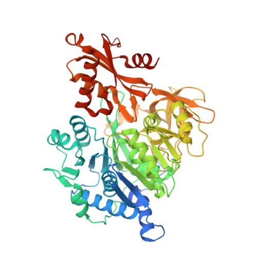
2WD9

CRYSTAL STRUCTURE OF HUMAN ACYL-COA SYNTHETASE MEDIUM-CHAIN FAMILY MEMBER 2A (L64P MUTATION) IN COMPLEX WITH IBUPROFEN


Experimental Data Snapshot

  • Method: X-RAY DIFFRACTION
  • Resolution: 2.60 Å
  • R-Value Free: 0.255 
  • R-Value Work: 0.235 
  • R-Value Observed: 0.236 

Starting Model: experimental
View more details

wwPDB Validation   3D Report Full Report


Ligand Structure Quality Assessment 


This is version 1.3 of the entry. See complete history


Literature

Structural Snapshots for the Conformation- Dependent Catalysis by Human Medium-Chain Acyl- Coenzyme a Synthetase Acsm2A.

Kochan, G.Pilka, E.S.Delft, F.V.Oppermann, U.Yue, W.W.

(2009) J Mol Biol 388: 997

  • DOI: https://doi.org/10.1016/j.jmb.2009.03.064
  • Primary Citation of Related Structures:  
    2VZE, 2WD9, 3B7W, 3C5E, 3DAY, 3EQ6, 3GPC

  • PubMed Abstract: 

    Acyl-CoA synthetases belong to the superfamily of adenylate-forming enzymes, and catalyze the two-step activation of fatty acids or carboxylate-containing xenobiotics. The carboxylate substrate first reacts with ATP to form an acyl-adenylate intermediate, which then reacts with CoA to produce an acyl-CoA ester. Here, we report the first crystal structure of a medium-chain acyl-CoA synthetase ACSM2A, in a series of substrate/product/cofactor complexes central to the catalytic mechanism. We observed a substantial rearrangement between the N- and C-terminal domains, driven purely by the identity of the bound ligand in the active site. Our structures allowed us to identify the presence or absence of the ATP pyrophosphates as the conformational switch, and elucidated new mechanistic details, including the role of invariant Lys557 and a divalent magnesium ion in coordinating the ATP pyrophosphates, as well as the involvement of a Gly-rich P-loop and the conserved Arg472-Glu365 salt bridge in the domain rearrangement.


  • Organizational Affiliation

    Structural Genomics Consortium, Old Road Research Campus Building, Oxford, UK.


Macromolecules
Find similar proteins by:  (by identity cutoff)  |  3D Structure
Entity ID: 1
MoleculeChains Sequence LengthOrganismDetailsImage
ACYL-COENZYME A SYNTHETASE ACSM2A, MITOCHONDRIAL
A, B, C
569Homo sapiensMutation(s): 1 
EC: 6.2.1.2 (PDB Primary Data), 6.2.1.25 (UniProt)
UniProt & NIH Common Fund Data Resources
Find proteins for Q08AH3 (Homo sapiens)
Explore Q08AH3 
Go to UniProtKB:  Q08AH3
PHAROS:  Q08AH3
GTEx:  ENSG00000183747 
Entity Groups  
Sequence Clusters30% Identity50% Identity70% Identity90% Identity95% Identity100% Identity
UniProt GroupQ08AH3
Sequence Annotations
Expand
  • Reference Sequence
Experimental Data & Validation

Experimental Data

  • Method: X-RAY DIFFRACTION
  • Resolution: 2.60 Å
  • R-Value Free: 0.255 
  • R-Value Work: 0.235 
  • R-Value Observed: 0.236 
  • Space Group: P 41 21 2
Unit Cell:
Length ( Å )Angle ( ˚ )
a = 98.32α = 90
b = 98.32β = 90
c = 381.44γ = 90
Software Package:
Software NamePurpose
REFMACrefinement
XDSdata reduction
XSCALEdata scaling
PHASERphasing

Structure Validation

View Full Validation Report


Ligand Structure Quality Assessment 


Entry History 

Deposition Data

Revision History  (Full details and data files)

  • Version 1.0: 2009-04-21
    Type: Initial release
  • Version 1.1: 2011-07-13
    Changes: Advisory, Version format compliance
  • Version 1.2: 2019-04-03
    Changes: Data collection, Other, Source and taxonomy
  • Version 1.3: 2023-12-13
    Changes: Data collection, Database references, Derived calculations, Other, Refinement description